Porcine epidemic diarrhea virus (PEDV) causes an emerging and re-emerging coronavirus disease characterized by vomiting, acute diarrhea, dehydration, and up to 100% mortality in neonatal suckling piglets, leading to huge economic losses in the global swine industry. Vaccination remains the most promising and effective way to prevent and control PEDV.
Porcine epidemic diarrhea virus (PEDV) was first reported in the United Kingdom. It is an enveloped virus with a single-stranded, positive-sense RNA genome that belongs to the genus Alphacoronavirus. The genome of PEDV is approximately 28 kb encoding 16 non-structural proteins (nsp1-nsp16), spike protein (S), envelope protein (E), membrane protein (M), nucleocapsid protein (N), etc.
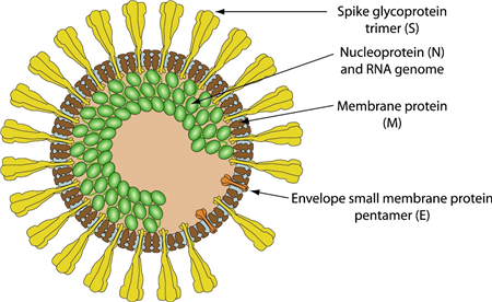
Figure 1. Schematic representation of PEDV (viralzone)
non-structural protein 1 (nsp1)
Nsp1 is recognized as a virulence factor in coronaviruses, playing a crucial role in the broad suppression of host gene expression to evade the innate immune response and promote viral replication. Structural analysis through crystallography has revealed that PEDV nsp1 is composed of two α-helices and six β-strands, with a six-stranded β-barrel structure centrally located between the two α-helices.
Papain-like protease (PLpro, nsp3)
Coronaviral papain-like proteases (PLpros) are essential enzymes that mediate not only the proteolytic processes of viral polyproteins during virus replication but also the deubiquitination and deISGylation of cellular proteins that attenuate host innate immune responses
Nsp3 encodes Papain-like protease (PLpro) present in various coronaviruses. The PLpro are responsible for the cleavage of the portion of the ORF1ab polyprotein encoded by the incoming RNA genome, which is essential for viral RNA synthesis [34]. The PLpro usually cleaves nsp1-4 and the cleavage sites can be summarized with LXGG↓ or the similar motif in SARS-CoV and MHV.
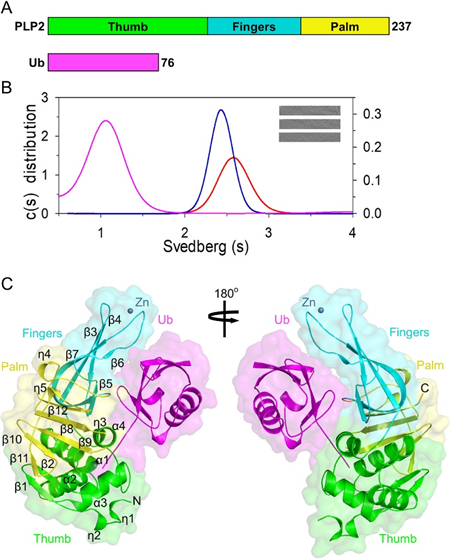
Figure 2. Structural characterization of the PEDV nsp3/Ub complex (Chu et al., 2022)
3C-like protease (3CLpro, nsp3)
3C-like protease (3CLpro), encoded by the gene for nsp5 which plays a pivotal role in coronaviruses replication, thus serving as an appealing antiviral drug target. Inhibition of PEDV nsp5 effectively inhibited the replication of PEDV, for instance, the broad-spectrum inhibitor GC376 reduced viral replication via binding the catalytic pocket of PEDV 3CLpro.
The 3C-like protease (3CLpro) is essential for the coronaviral life cycle and is an appealing target for the development of therapeutics.
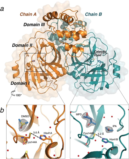
Figure 3. Crystal structure of PEDV 3CLpro (St. John et al., 2016)
Spike protein
The S protein, a type I glycoprotein, is essential for viral entry into host cells and consists of two subunits: S1 and S2. The S1 subunit includes an N-terminal signal peptide, a sialic acid binding region (19-233 aa), and a core neutralizing epitope (499-638 aa). It facilitates attachment to host cells by binding to receptors such as ANPEP (aminopeptidase N), thereby initiating the infection process. This receptor interaction likely induces conformational changes that unmask the fusion peptide located within the S2 subunit. The S2 subunit comprises a fusion peptide (891-908 aa), two heptad repeat regions (HR1: 978-1117 aa; HR2: 1274-1313 aa), a transmembrane domain (1328-1350 aa), and a cytoplasmic domain (1351-1386 aa).
The S protein is proposed to exist in at least three conformational states: pre-fusion native state, pre-hairpin intermediate state, and post-fusion hairpin state. During the fusion process, the heptad repeat regions rearrange to form a trimer-of-hairpins structure, positioning the fusion peptide adjacent to the C-terminal region of the ectodomain. This conformational transition is thought to facilitate the apposition and subsequent fusion of the viral and target cell membranes, ultimately enabling viral entry.
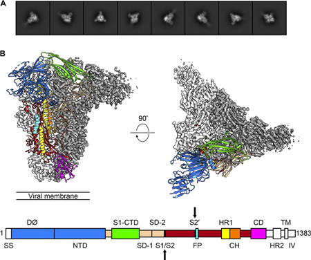
Figure 4. Cryo-EM Structure of the PEDV Spike Protein in the Prefusion Conformation (Wrapp and McLellan, 2019)
N protein
The nucleocapsid (N) protein of porcine epidemic diarrhea virus (PEDV) is a highly conserved protein that plays a critical role in viral replication and transcription. It forms a complex with the viral RNA genome, serving as the core of the virus. The N protein packages the positive-strand viral genome RNA into a helical ribonucleocapsid (RNP) and is fundamental in virion assembly through its interactions with the viral genome and the membrane protein M.
The N protein is multifunctional; as a structural component, it contributes to nucleocapsid formation and enhances the efficiency of subgenomic RNA transcription and overall viral replication. It is often phosphorylated, particularly in the N-terminal domain and the serine-arginine-rich (SR) region, modifications that are crucial for RNA binding and replication efficiency. Additionally, the C-terminal domain facilitates dimerization of the N protein, further supporting its roles in viral biology.
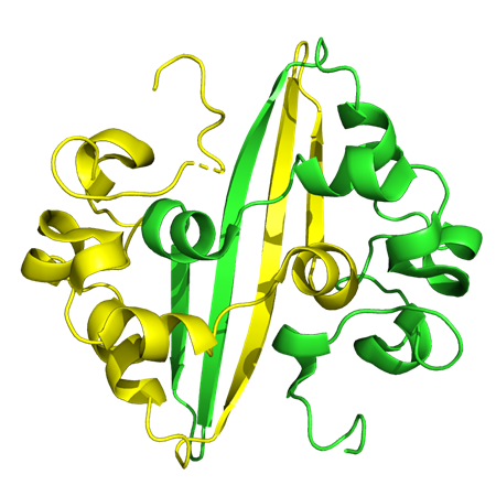
Figure 5. Crystal structure of the C-terminal domain of PEDV nucleocapsid protein
Reference
1. Chu, H.-F., Cheng, S.-C., Sun, C.-Y., Chou, C.-Y., Lin, T.-H., and Chen, W.-Y. (2022). Structural and Biochemical Characterization of Porcine Epidemic Diarrhea Virus Papain-Like Protease 2. Journal of Virology 96, e01372-01321.
2. Fredmoore, L.O. (2023). Immune evasion mechanisms of porcine epidemic diarrhea virus: A comprehensive review: https://doi.org/10.12982/VIS.2024.014. Veterinary Integrative Sciences 22, 171-192.
3. Hu, Y., Xie, X., Yang, L., and Wang, A. (2021). A Comprehensive View on the Host Factors and Viral Proteins Associated With Porcine Epidemic Diarrhea Virus Infection. Front Microbiol 12, 762358.
4. Li, Z., Ma, Z., Li, Y., Gao, S., and Xiao, S. (2020). Porcine epidemic diarrhea virus: Molecular mechanisms of attenuation and vaccines. Microbial Pathogenesis 149, 104553.
5. Lin, F., Zhang, H., Li, L., Yang, Y., Zou, X., Chen, J., and Tang, X. (2022). PEDV: Insights and Advances into Types, Function, Structure, and Receptor Recognition. Viruses 14, 1744.
6. Luo, H., Liang, Z., Lin, J., Wang, Y., Liu, Y., Mei, K., Zhao, M., and Huang, S. (2024). Research progress of porcine epidemic diarrhea virus S protein. Front Microbiol 15, 1396894.
7. St. John, S.E., Anson, B.J., and Mesecar, A.D. (2016). X-Ray Structure and Inhibition of 3C-like Protease from Porcine Epidemic Diarrhea Virus. Scientific Reports 6, 25961.
8. Wrapp, D., and McLellan, J.S. (2019). The 3.1-Angstrom Cryo-electron Microscopy Structure of the Porcine Epidemic Diarrhea Virus Spike Protein in the Prefusion Conformation. Journal of Virology 93, 10.1128/jvi.00923-00919.
9. Zhang, Y., Chen, Y., Zhou, J., Wang, X., Ma, L., Li, J., Yang, L., Yuan, H., Pang, D., and Ouyang, H. (2022). Porcine Epidemic Diarrhea Virus: An Updated Overview of Virus Epidemiology, Virulence Variation Patterns and Virus–Host Interactions. Viruses 14, 2434
Host species: Rabbit
Isotype: IgG
Applications: ELISA, IHC, WB
Accession: S0AP48
Host species: Rabbit
Isotype: IgG
Applications: ELISA, IHC, WB
Accession: S0AP48
Host species: Rabbit
Isotype: IgG
Applications: ELISA, IHC, WB
Accession: ALS35468.1
Applications: ELISA, Immunogen, SDS-PAGE, WB, Bioactivity testing in progress
Expression system: E. coli
Accession: ALS35468.1
Protein length: Ala2990-Gln3291