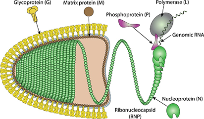
Fig. 1 Schematic representation of Chandipura virus (viralzone)
Chandipura virus (CHPV; Family Rhabdoviridae, Genus Vesiculovirus) is an emerging human pathogen associated with deadly encephalitis, principally among children in the tropical areas of India. It predominantly affects children and is characterized by influenza-like illness and neurologic dysfunctions. It is transmitted by vectors such as mosquitoes, ticks and sand flies. Currently, there is no vaccine or specific antiviral treatment for CHPV infection.
Chandipura virus was discovered in 1966 by Bhatt and Rodriguez, at Virus Research Centre (VRC), Pune. It was found accidentally while investigating patients suffering from fever in Chandipura village in northern Maharashtra near Nagpur district, for dengue or chikungunya virus etiology, and was thus named Chandipura Virus.
CHPV is surrounded by lipoprotein envelope, which encloses a helical ribonucleoparticle (RNP) with a non-segmented single strand RNA. The genome is about 11000 nucleotides encoding five different proteins Nucleoprotein, Phosphoprotein, Matrix protein, Glycoprotein and RNA-directed RNA polymerase L.
Nucleoprotein
The viral genomic RNA is encapsidated by the nucleocapsid protein (N) into a helical structure, a process essential for protecting the genome from cellular ribonucleases and enabling its function as a template for viral RNA synthesis. In rhabdoviruses, nucleocapsid assembly occurs concomitantly with genome replication, with encapsidation relying on both N–N protein interactions and N–RNA contacts. When expressed in isolation, the N protein tends to aggregate and binds non-viral RNA indiscriminately. The Chandipura virus (CHPV) N protein exists in two distinct forms: a higher-order oligomeric form and a monomeric form. In its oligomeric state, N exhibits broad RNA-binding specificity, interacting with both viral and non-viral RNAs, whereas the monomeric form specifically recognizes sequence elements in the viral leader RNA, a critical determinant for precise viral genome encapsidation. The phosphoprotein (P) prevents the oligomerization of CHPV N, maintaining it in a monomeric form. This regulation is crucial for imparting RNA-binding specificity, as monomeric N selectively interacts with viral RNA rather than non-viral RNAs. This mechanism highlights the role of P in modulating the functionality of N during viral genome replication and encapsidation.
Matrix protein
The matrix protein (M) is critical for rhabdoviral structure, assembly, and pathogenicity. Positioned on the inner surface of the virion, M connects the nucleocapsid to the membrane via its highly basic N-terminal domain, which includes eight lysine residues and a conserved triple proline sequence. Structural studies of vesicular stomatitis virus (VSV) M protein highlight additional contributions from a secondary domain and a flexible linker conserved among mononegavirales, ensuring proper membrane alignment.
M protein plays a key role in viral assembly and budding by recruiting mature nucleocapsid particles to the membrane and condensing the ribonucleocapsid core. It also inhibits viral transcription, likely by inducing a condensed conformation of the ribonucleoprotein complex (RNP). Beyond its structural functions, M suppresses host gene expression by inhibiting mRNA nuclear export through interaction with the RAE1-NUP98 complex, thereby blocking interferon signaling and antiviral responses. Additionally, M induces cell rounding, cytoskeletal disorganization, and apoptosis, contributing to the cytotoxicity observed in rhabdovirus-infected cells.
Glycoprotein
The entry of Chandipura virus (CHPV) into susceptible cells is mediated by its single envelope glycoprotein G, which performs dual roles in receptor recognition and membrane fusion. Initially, glycoprotein G facilitates viral attachment to the host cell by interacting with a cellular receptor, triggering endocytosis of the virion. For vesicular stomatitis virus Indiana (VSVIND), a closely related virus, the receptor has been identified as the LDL receptor and its family members.
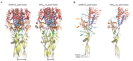
Following endocytosis, the acidic environment of the endosome induces conformational changes in glycoprotein G. Specifically, G transitions from its trimeric pre-fusion (PRE) conformation to its trimeric post-fusion (POST) state. This structural rearrangement drives the fusion of the viral and endosomal membranes, allowing the release of the viral nucleocapsid into the cytoplasm and initiating subsequent steps of infection. Thus, glycoprotein G is critical for two key stages of CHPV entry: receptor recognition and pH-dependent membrane fusion.

Fig.2 Structure of CHPV-Gth and VSVIND-Gth post-fusion trimers (Baquero et al., 2015)
Reference
1. Baquero, E., Albertini, A.A., Raux, H., Abou‐Hamdan, A., Boeri‐Erba, E., Ouldali, M., Buonocore, L., Rose, J.K., Lepault, J., Bressanelli, S., et al. (2017). Structural intermediates in the fusion‐associated transition of vesiculovirus glycoprotein. The EMBO Journal 36, 679-692.
2. Baquero, E., Albertini, A.A., Raux, H., Buonocore, L., Rose, J.K., Bressanelli, S., and Gaudin, Y. (2015). Structure of the Low pH Conformation of Chandipura Virus G Reveals Important Features in the Evolution of the Vesiculovirus Glycoprotein. PLOS Pathogens 11, e1004756.
3. Menghani, S., Chikhale, R., Raval, A., Wadibhasme, P., and Khedekar, P. (2012). Chandipura Virus: An emerging tropical pathogen. Acta Tropica 124, 1-14.
4. Mondal, A., Bhattacharya, R., Ganguly, T., Mukhopadhyay, S., Basu, A., Basak, S., and Chattopadhyay, D. (2010). Elucidation of functional domains of Chandipura virus Nucleocapsid protein involved in oligomerization and RNA binding: Implication in viral genome encapsidation. Virology 407, 33-42.
Host species: Rabbit
Isotype: IgG
Applications: ELISA, IHC, WB
Accession: P11211
Host species: Rabbit
Isotype: IgG
Applications: ELISA, IHC, WB
Accession: Q9WH76
Host species: Rabbit
Isotype: IgG
Applications: ELISA, IHC, WB
Accession: P16380
Host species: Rabbit
Isotype: IgG
Applications: ELISA, IHC, WB
Accession: P13180
Applications: ELISA, Immunogen, SDS-PAGE, WB, Bioactivity testing in progress
Expression system: E. coli
Accession: P11211
Protein length: Met1-Ala422
Applications: ELISA, Immunogen, SDS-PAGE, WB, Bioactivity testing in progress
Expression system: E. coli
Accession: Q9WH76
Protein length: Phe47-His229
Applications: ELISA, Immunogen, SDS-PAGE, WB, Bioactivity testing in progress
Expression system: E. coli
Accession: P16380
Protein length: Thr104-Asn293
Applications: ELISA, Immunogen, SDS-PAGE, WB, Bioactivity testing in progress
Expression system: E. coli
Accession: P13180
Protein length: Tyr22-Ser440
Applications: ELISA, Immunogen, SDS-PAGE, WB, Bioactivity testing in progress
Expression system: Mammalian cells
Accession: P13180
Protein length: Tyr22-Ser440
Applications: ELISA, Immunogen, SDS-PAGE, WB, Bioactivity testing in progress
Expression system: Mammalian cells
Accession: P13180
Protein length: Tyr22-Ser440