Foot-and-mouth disease virus (FMDV), the prototype of the Aphthovirus genus, is a small, non-enveloped virus with a single-stranded, positive-sense RNA genome of approximately 8,400 nucleotides. It primarily infects cloven-hoofed animals, causing foot-and-mouth disease (FMD), a highly contagious and acute illness. FMD leads to severe economic losses, including decreased milk production in dairy cattle and reduced growth rates in meat-producing animals. Despite control measures, the disease remains widespread globally, posing a significant threat to livestock and agricultural economies.
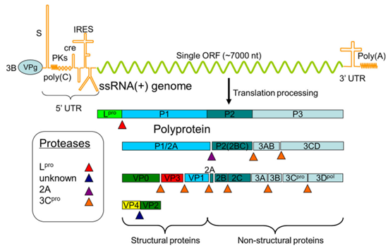
The genome of FMDV encodes a polyprotein that undergoes successive cleavage by viral and cellular proteases, resulting in the production of mature structural proteins (VP1, VP2, VP3, and VP4), which form the viral capsid, and non-structural proteins (Lpro, 2A, 2B, 2C, 3A, 3B, 3C, and 3D), which primarily function in viral replication.

Fig. 1 Schematic map of the FMDV genome showing the encoded proteins and the proteolytic reaction cascade producing the final products. (Han et al., 2014)
VP proteins
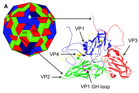
The viral capsid of FMDV is composed of 60 copies each of four viral-encoded proteins: VP1, VP2, VP3, and VP4. Among these, VP1, VP2, and VP3 form the outer capsid shell, while VP4 constitutes the interior surface. The VP1 protein consists of 213 amino acids, with most of these residues located around the five-fold axis, forming multiple exposed loops in this region. It has been suggested that the flexibility of the GH loop in VP1 plays a crucial role in facilitating the virus's binding to antibodies and cell receptors. VP2 and VP3 are alternately arranged around the three-fold axis of the capsid. The N-terminus of VP4 is positioned near the five-fold axis, while its C-terminus is located near the three-fold axis.

Fig. 2 Conformations of the structural proteins (Han et al., 2014)
Lpro
Protein synthesis on the FMDV genome can initiate at one of two in-frame AUG codons, which are 84 nucleotides apart. As a result, Lpro can exist in two isoforms, Labpro and Lbpro, differing by 28 amino acids. Lpro is a papain-like cysteine protease that autocatalytically cleaves itself from the polyprotein at the L/VP0 junction. It also cleaves host translation initiation factors EIF4G1 and EIF4G3, thereby inhibiting the translation of capped cellular mRNAs. In addition, Lpro contributes to counteracting the host's innate antiviral response through various mechanisms.
2C Protein
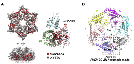
Protein 2C is one of the most highly conserved viral proteins encoded by FMDV and belongs to the 2C-like (2CL) protein family, which is found in many other RNA viruses across animals and plants. 2C associates with and induces structural rearrangements of intracellular membranes. By interacting with the host protein BECN1, it triggers autophagy, promoting viral replication. Protein 2C forms a homohexamer and also interacts with the host protein VIM.

Fig. 3 Hexameric ring model of FMDV 2C (Zhang et al., 2022)
3C Protein
The FMDV 3C proteinase, responsible for most of the cleavages in the viral polyprotein, has been identified as a chymotrypsin-like cysteine protease. This protease generates mature viral proteins from the precursor polyprotein. In addition to its proteolytic activity, 3C binds to viral RNA, influencing viral genome replication. Both RNA and substrate bind cooperatively to the protease, enhancing its activity.
Reference
1. Cencic, R., Mayer, C., Juliano, M.A., Juliano, L., Konrat, R., Kontaxis, G., and Skern, T. (2007). Investigating the substrate specificity and oligomerisation of the leader protease of foot and mouth disease virus using NMR. J Mol Biol 373, 1071-1087.
2. Gao, Y., Sun, S.Q., and Guo, H.C. (2016). Biological function of Foot-and-mouth disease virus non-structural proteins and non-coding elements. Virol J 13, 107.
3. Grubman, M.J., and Baxt, B. (2004). Foot-and-Mouth Disease. Clinical Microbiology Reviews 17, 465-493.
4. Han, S.-C., Guo, H.-C., and Sun, S.-Q. (2014). Three-dimensional structure of foot-and-mouth disease virus and its biological functions. Archives of Virology 160, 1-16.
5. Ruiz-Sáenz, J., Goez, Y., Tabares, W., and López-Herrera, A. (2009). Cellular Receptors for Foot and Mouth Disease Virus. Intervirology 52, 201-212.
6. Zhang, C., Yang, F., Wojdyla, J.A., Qin, B., Zhang, W., Zheng, M., Cao, W., Wang, M., Gao, X., Zheng, H., et al. (2022). An anti-picornaviral strategy based on the crystal structure of foot-and-mouth disease virus 2C protein. Cell Reports 40.
7. Zunszain, P.A., Knox, S.R., Sweeney, T.R., Yang, J., Roque-Rosell, N., Belsham, G.J., Leatherbarrow, R.J., and Curry, S. (2010). Insights into cleavage specificity from the crystal structure of foot-and-mouth disease virus 3C protease complexed with a peptide substrate. J Mol Biol 395, 375-389.
Host species: Rabbit
Isotype: IgG
Applications: ELISA, IHC, WB
Accession: ADV38199.1
Applications: ELISA, Immunogen, SDS-PAGE, WB, Bioactivity testing in progress
Expression system: E. coli
Accession: ADV38199.1
Protein length: Thr1-Gln211
Host species: Human
Isotype: IgG2a, kappa
Applications: ELISA, WB
Accession: P03306
Host species: Human
Isotype: IgG1, kappa
Applications: IHC, Neutralization
Accession: P03309, Q6PMW3
Host species: Human
Isotype: IgG1, kappa
Applications: IHC, Neutralization, WB
Accession: P03309, Q6PMW3
Host species: Human
Isotype: IgG1, kappa
Applications: ELISA, WB
Accession: P03309
Host species: Human
Isotype: IgG1, kappa
Applications: ELISA, WB
Accession: P03309
Host species: Human
Isotype: IgG1, kappa
Applications: ELISA
Accession: P15072
Host species: Human
Isotype: IgG1, kappa
Applications: ELISA, WB
Accession: P03311