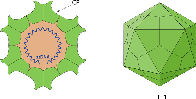
Fig 1. Schematic representation of Adenoviruse (viralzone)
Parvovirus B19 (B19V), a small single-stranded DNA virus belonging to the family Parvoviridae, subfamily Parvovirinae, and genus Erythrovirus, was first identified in 1975 and linked to human disease in 1981. B19V is responsible for various clinical syndromes, including fifth disease, transient aplastic crisis, pure red cell aplasia, hydrops fetalis, glomerulopathy, and anemia in end-stage renal disease. The virus is primarily acquired during childhood, with serological evidence of past infection found in 70-85% of adults, demonstrating seasonal variation in infectivity, particularly in winter and spring. Transmission occurs mainly via inhalation of aerosolized droplets, with additional routes including vertical transmission from mother to fetus, and through blood product transfusions, bone marrow, and solid-organ transplants. The adaptive immune response to B19V is characterized by the early production of IgM antibodies, which typically persist for 3-6 months post-infection, followed by the development of long-lasting IgG antibodies, with IgA also detectable in various body fluids. Currently, there is no approved vaccine for B19, and B19 infection is still a threat in certain circumstances.
The genome of Parvovirus B19 (B19V) is approximately 5.6 kb in length and encodes six proteins: the viral capsid proteins VP1 and VP2, the main replication protein NS1, and three smaller nonstructural proteins. The single-stranded DNA genome is encapsulated within a nonenveloped, icosahedral protein shell measuring 20 nm in diameter. This capsid comprises 60 structural subunits, with about 95% being the major viral protein VP2 (58 kDa). VP1, the other structural protein, differs from VP2 by an N-terminal "unique region" containing 227 additional amino acids, primarily located outside the virion, thus making it more accessible for antibody binding.
NS1 (Q9PZT1)
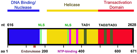
NS1 is a multifunctional protein essential for the replication of the B19 virus (B19V) genome. This 671 amino acid protein, with a molecular weight of approximately 78 kDa, possesses several important domains. The N-terminus (amino acids 2-176) harbors DNA binding and endonuclease activity, with a specific endonuclease motif located between amino acids 137 and 145. This endonuclease domain is crucial for terminal resolution and supports rolling hairpin replication, facilitating viral genome replication. Additionally, NS1 features two nuclear localization signals, KKPR (177-179) and KKCGKK (316-321), which direct its predominant localization to the nucleus of infected cells. The central region of NS1 contains ATPase and NTP-binding domains, while the C-terminus includes a transactivation domain. NS1 interacts cooperatively with the viral DNA origin of replication and transactivates various promoters, including the viral p6 promoter, underscoring its vital role during B19V infection.

Fig 2. A diagram of NS1 functional domains. (Ganaie and Qiu, 2018)
Capsid Proteins (VP1 and VP2)
VP1 is a minor capsid protein, consisting of 781 amino acids (~84 kDa), which shares a common C-terminus with the major capsid protein VP2, with the addition of 227 unique amino acids termed the VP1-unique (VP1u) region. VP2, with a length of 554 amino acids (~58 kDa), possesses a nuclear localization signal at its C-terminus, resulting in the localization of both VP1 and VP2 within the nucleus of infected cells. VP1 is expressed at lower levels and assembles with VP2 into the viral capsid at a ratio of 1:20. Initial interactions occur between the capsid and the P antigen, with the first 100 amino acids of VP1u facilitating the internalization of virus particles. Notably, the VP1u region (amino acids 128 to 160) exhibits phospholipase A2 activity, which is proposed to assist in evading lysosomal fusion and promote the nuclear entry of the virions.
The capsid self-assembles into an icosahedral structure with T=1 symmetry, approximately 20 nm in diameter, comprising 60 copies of the two capsid protein variants, VP1 and VP2. This capsid encapsulates the genomic ssDNA and interacts with erythroid progenitor cells expressing high levels of P antigen, utilizing host ITGA5-ITGB1 and XRCC5/Ku80 autoantigens as coreceptors to mediate virion attachment. This interaction induces virion internalization primarily through clathrin-dependent endocytosis. Moreover, binding to host receptors prompts capsid rearrangements that expose the N-terminus of VP1, particularly its phospholipase A2-like region. The additional N-terminal extension of VP1u may function as a lipolytic enzyme, facilitating the breach of the endosomal membrane during entry and potentially contributing to viral transport to the nucleus. In summary, while the structural proteins VP1 and VP2 are responsible for forming the viral capsid for DNA encapsidation, non-structural proteins play critical roles in viral replication, packaging, and the release of infectious particles, necessitating further investigation into their functional characterization.

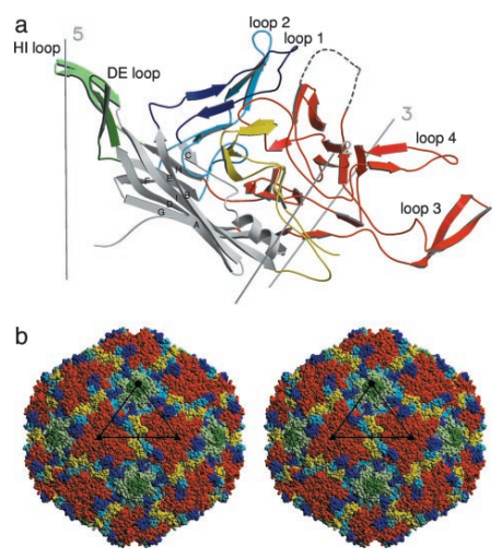
Fig 3. Secondary structure of parvovirus B19 (Kaufmann et al., 2004)
Reference
1. Dittmer, F.P., Guimaraes, C.M., Peixoto, A.B., Pontes, K.F.M., Bonasoni, M.P., Tonni, G., and Araujo Junior, E. (2024). Parvovirus B19 Infection and Pregnancy: Review of the Current Knowledge. J Pers Med 14.
2. Farahmand, M., Tavakoli, A., Ghorbani, S., Monavari, S.H., Kiani, S.J., and Minaeian, S. (2021). Molecular and serological markers of human parvovirus B19 infection in blood donors: A systematic review and meta-analysis. Asian J Transfus Sci 15, 212-222.
3. Ganaie, S.S., and Qiu, J. (2018). Recent Advances in Replication and Infection of Human Parvovirus B19. Front Cell Infect Microbiol 8, 166.
4. Kaufmann, B., Simpson, A.A., and Rossmann, M.G. (2004). The structure of human parvovirus B19. Proc Natl Acad Sci U S A 101, 11628-11633.
5. Rajendiran, P., Saravanan, N., Ramamurthy, M., Nandagopal, B., and Vadivel, K. (2022). An Overview of the Epidemiology, Pathogenesis, Diagnosis, and Treatment of Human Parvovirus B19. Asian Journal of Research in Infectious Diseases, 32-43.
6. Rogo, L.D., Mokhtari-Azad, T., Kabir, M.H., and Rezaei, F. (2014). Human parvovirus B19: a review. Acta Virol 58, 199-213.
7. Sanchez, J.L., Ghadirian, N., and Horton, N.C. (2022). High-Resolution Structure of the Nuclease Domain of the Human Parvovirus B19 Main Replication Protein NS1. J Virol 96, e0216421.
8. Sun, Y., Klose, T., Liu, Y., Modrow, S., and Rossmann, M.G. (2019). Structure of Parvovirus B19 Decorated by Fabs from a Human Antibody. J Virol 93.
9. Zhang, Y., Shao, Z., Gao, Y., Fan, B., Yang, J., Chen, X., Zhao, X., Shao, Q., Zhang, W., Cao, C., et al. (2022). Structures and implications of the nuclease domain of human parvovirus B19 NS1 protein. Comput Struct Biotechnol J 20, 4645-4655.
Host species: Rabbit
Isotype: IgG
Applications: ELISA, IHC, WB
Accession: YP_004928146.1
Host species: Rabbit
Isotype: IgG
Applications: ELISA, IHC, WB
Accession: YP_004928148.1
Host species: Rabbit
Isotype: IgG
Applications: ELISA, IHC, WB
Accession: Q6TV13
Applications: ELISA, Immunogen, SDS-PAGE, WB, Bioactivity testing in progress
Expression system: E. coli
Accession: YP_004928146.1
Protein length: His80-Ser227
Applications: ELISA, Immunogen, SDS-PAGE, WB, Bioactivity testing in progress
Expression system: E. coli
Accession: YP_004928148.1
Protein length: Val21-Phe234
Applications: ELISA, Immunogen, SDS-PAGE, WB, Bioactivity testing in progress
Expression system: E. coli
Accession: Q6TV13
Protein length: Met1-Glu671
Host species: Mouse
Isotype: IgG1, kappa
Applications: Neutralization
Accession: P08508, P08101