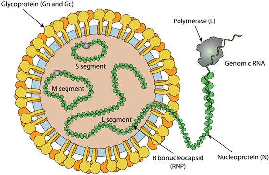
Figure 1 Schematic representation of Hantaan virus (viralzone)
Hantaviruses belong to the genus Orthohantavirus, a group of single-stranded, enveloped, negative-sense RNA viruses within the family Hantaviridae, part of the order Bunyavirales. These viruses are named after the Hantan River in South Korea, where the first member, Hantaan virus, was isolated in 1976. These viruses are primarily associated with rodents and can cause severe diseases in humans upon zoonotic transmission.
Orthohantaviruses typically cause chronic asymptomatic infections in rodents, which shed the virus in their urine, saliva, and feces. Human infection occurs through direct contact with contaminated materials or by inhalation of aerosolized virus particles. This transmission route underscores the importance of rodent control in preventing outbreaks.
In humans, hantaviruses can lead to severe diseases such as Hantavirus Pulmonary Syndrome (HPS). HPS is one of two potentially fatal syndromes of zoonotic origin caused by species of hantavirus. These include Black Creek Canal virus (BCCV), New York orthohantavirus (NYV), Monongahela virus (MGLV), Sin Nombre orthohantavirus (SNV), and certain other members of hantavirus genera that are native to the United States and Canada.
While most hantaviruses are transmitted through rodents, instances of human-to-human transmission have been documented. Notably, the Andes virus has been reported to spread between individuals in South America, although such cases are rare.
The hantavirus virion is spherical or tubular, with sizes ranging widely from 70 to 350 nm in diameter or length. It consists of RNA segments encapsulated by the nucleocapsid protein (N) to form ribonucleoprotein (RNP). The lipid envelope is studded with glycoprotein spikes, which are crucial for viral entry into host cells.
Orthohantaviruses are characterized by their RNA genome segmented into small (S), medium (M), and large (L) segments. They are enveloped particles with glycoprotein spikes (Gn and Gc) protruding from their lipid envelope.
Research on hantaviruses continues to uncover new species and strains, improving our understanding of their epidemiology and potential treatments. Challenges include developing effective vaccines and antiviral therapies due to the diversity and adaptability of hantaviruses.
Envelopment polyprotein (P0DTJ0)
Glycoprotein N:
Glycoprotein N (Gn, also known as G1) is produced by enzymatic cleavage of the glycoprotein precursor (GPC) protein encoded in the M segment and are jointly responsible for orchestrating host cell entry and fusion. The maturing Gn is decorated by N-linked glycans that are thought to be involved in protein folding, receptor binding, membrane fusion and viral morphogenesis. Glycoprotein N forms homotetramers with glycoprotein C at the surface of the virion.
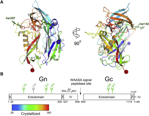
Figure 2 Crystal structure of Gn (Li et al., 2016)
Glycoprotein C:
Glycoprotein C (Gc, also konw as G2) forms homotetramers with glycoprotein N at the surface of the virion. Attaches the virion to host cell receptors including integrin ITGAV/ITGB3. This attachment induces virion internalization predominantly through clathrin-dependent endocytosis. Class II fusion protein that promotes fusion of viral membrane with host endosomal membrane after endocytosis of the virion.
Antibodies that bind Gn and Gc have neutralizing activity and provide lasting protection in vivo. They are two important targets to develop effective vaccines and generate neutralizing antibodies.
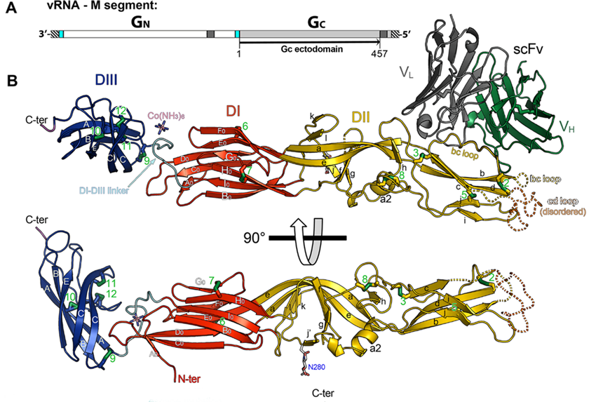
Figure 3 Structure of Hantaan virus Gc (Guardado-Calvo et al., 2016)
RNA-directed RNA polymerase L (V5IVB1)
RdRp which responsible for the replication and transcription of the viral RNA genome, is the largest protein encoded by hantaviruses which derived from the L segment. By mobility in SDS-PAGE the molecular mass of RdRp hasbeen estimated to be ~250 kDa. The RdRp of hantaviruses is capable of recombining homologous RNA sequences and thus enables virus evolution via superinfection. The RdRp of hantaviruses requires a divalent cation (either Mg2+ or Mn2+) for activity with a preference for Mn2+. During transcription, synthesizes subgenomic RNAs and assures their capping by a cap-snatching mechanism, which involves the endonuclease activity cleaving the host capped pre-mRNAs. These short capped RNAs are then used as primers for viral transcription. Cleaves ssRNA substrates but not DNA.Seems to downregulate the expression of its own and heterologous mRNAs through its endonuclease activity.
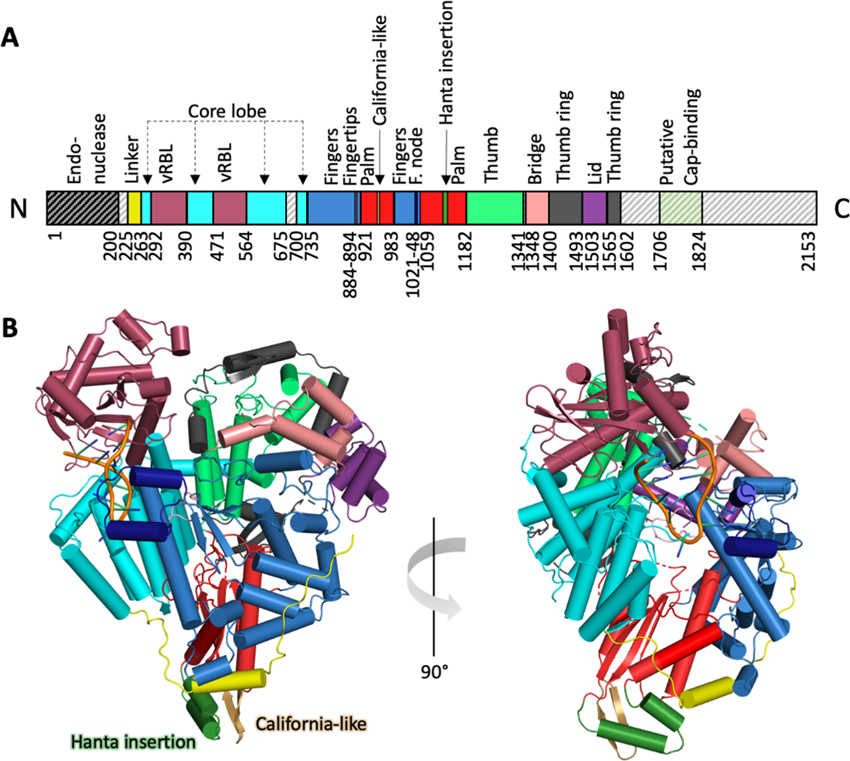
Figure 4 Structural and functional characterization of RdRp (Meier et al., 2023)
Nucleoprotein (I7GVL4)
The S segment-encoded N protein is a non-glycosylated protein with a molecular mass of approximately 50 kDa. Both the N protein and RdRp are required for replication. Thus, to provide a sufficient pool of the N protein, mRNA transcription and translation are believed to precede the initiation of replication and the concentration of free N protein is thought to mediate the switch from mRNA synthesis towards replication. Antibodies targeting the nucleoprotein (N) arise rapidly following infection, rendering them a useful biomarker for infection.
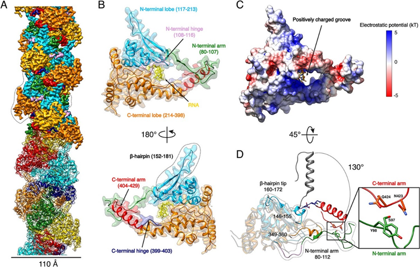
Figure 5 Structure of Nucleoprotein (Arragain et al., 2019)
Reference:
1. Arragain, B., Reguera, J., Desfosses, A., Gutsche, I., Schoehn, G., and Malet, H. (2019). High resolution cryo-EM structure of the helical RNA-bound Hantaan virus nucleocapsid reveals its assembly mechanisms. eLife 8, e43075.
2. Guardado-Calvo, P., Bignon, E.A., Stettner, E., Jeffers, S.A., Pérez-Vargas, J., Pehau-Arnaudet, G., Tortorici, M.A., Jestin, J.-L., England, P., Tischler, N.D., et al. (2016). Mechanistic Insight into Bunyavirus-Induced Membrane Fusion from Structure-Function Analyses of the Hantavirus Envelope Glycoprotein Gc. PLOS Pathogens 12, e1005813.
3. Hepojoki, J., Strandin, T., Lankinen, H., and Vaheri, A. (2012). Hantavirus structure – molecular interactions behind the scene. Journal of General Virology 93, 1631-1644.
4. Li, S., Rissanen, I., Zeltina, A., Hepojoki, J., Raghwani, J., Harlos, K., Pybus, O.G., Huiskonen, J.T., and Bowden, T.A. (2016). A Molecular-Level Account of the Antigenic Hantaviral Surface. Cell Reports 15, 959-967.
5. Meier, K., Thorkelsson, S.R., Durieux Trouilleton, Q., Vogel, D., Yu, D., Kosinski, J., Cusack, S., Malet, H., Grünewald, K., Quemin, E.R.J., et al. (2023). Structural and functional characterization of the Sin Nombre virus L protein. PLOS Pathogens 19, e1011533.
6. Rissanen, I., Krumm, S.A., Stass, R., Whitaker, A., Voss, J.E., Bruce, E.A., Rothenberger, S., Kunz, S., Burton, D.R., Huiskonen, J.T., et al. (2021). Structural Basis for a Neutralizing Antibody Response Elicited by a Recombinant Hantaan Virus Gn Immunogen. mBio 12.
7. Serris, A., Stass, R., Bignon, E.A., Muena, N.A., Manuguerra, J.-C., Jangra, R.K., Li, S., Chandran, K., Tischler, N.D., Huiskonen, J.T., et al. (2020). The Hantavirus Surface Glycoprotein Lattice and Its Fusion Control Mechanism. Cell 183, 442-456.e416.
Applications: ELISA, Immunogen, SDS-PAGE, WB, Bioactivity testing in progress
Expression system: E. coli
Accession: P05133
Protein length: Thr3-Arg93
Host species: Rabbit
Isotype: IgG
Applications: ELISA, IHC, WB
Accession: I7GVL4
Host species: Rabbit
Isotype: IgG
Applications: ELISA, IHC, WB
Accession: P0DTJ0
Host species: Rabbit
Isotype: IgG
Applications: ELISA, IHC, WB
Accession: P0DTJ0
Host species: Human
Isotype: IgG1
Applications: Research Grade Biosimilar
Expression system: Mammalian Cells
Host species: Human
Isotype: IgG1
Applications: Research Grade Biosimilar
Expression system: Mammalian Cells
Host species: Human
Isotype: IgG1
Applications: Research Grade Biosimilar
Expression system: Mammalian Cells
Host species: Human
Isotype: IgG1
Applications: ELISA, FCM, IF, WB
Accession: P08668