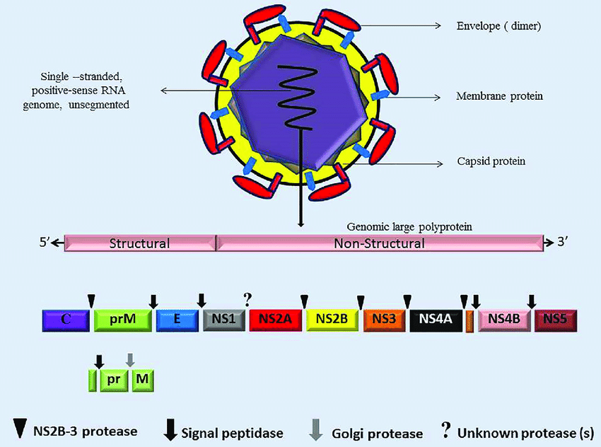
(Javed F, Manzoor KN, Ali M, et al. Zika virus: What we need to know? J Basic Microbiol. 2017;1–14.)
Zika virus (ZIKV)
Zika virus (ZIKV) is an Arbovirus first time discovered in 1947 from the forest of Uganda named Zika. Different outbreaks of Zika virus disease have been recorded in Africa, the Americas, Asia and the Pacific. Its infection is caused by the bite of an infected Aedes mosquito, usually causing mild fever, rash, conjunctivitis, and muscle pain.
Zika virus (ZIKV) belongs to the genus Flavivirus and family Flaviviridae. The diameter of the virion is approximately is 50–60 nm while its nucleocapsid arranged in icosahedral symmetry [47,48]. The virion includes a positive-sense ssRNA genome which contains one single open reading frame with untranslated regions (UTR) at 3′ and 5′ ends [46]. The Polypeptide protein of ZIKV is divided into three structural and seven non-structural proteins.
Genome polyprotein (Q32ZE1)
Function:
Capsid protein C:
Plays a role in virus budding by binding to the host cell membrane and packages the viral RNA into a nucleocapsid that forms the core of the mature virus particle. During virus entry, may induce genome penetration into the host cytoplasm after hemifusion induced by the surface proteins. Can migrate to the cell nucleus where it modulates host functions.
Inhibits RNA silencing by interfering with host Dicer.
Peptide pr:
Prevents premature fusion activity of envelope proteins in trans-Golgi by binding to envelope protein E at pH 6.0. After virion release in extracellular space, gets dissociated from E dimers.
Protein prM:
Plays a role in host immune defense modulation and protection of envelope protein E during virion synthesis. PrM-E cleavage is inefficient, many virions are only partially matured and immature prM-E proteins could play a role in immune evasion. Contributes to fetal microcephaly in humans. Acts as a chaperone for envelope protein E during intracellular virion assembly by masking and inactivating envelope protein E fusion peptide. prM is the only viral peptide matured by host furin in the trans-Golgi network probably to avoid catastrophic activation of the viral fusion activity in acidic Golgi compartment prior to virion release.
Small envelope protein M:
May play a role in virus budding. Exerts cytotoxic effects by activating a mitochondrial apoptotic pathway through M ectodomain. May display a viroporin activity.
Envelope protein E:
Binds to host cell surface receptors and mediates fusion between viral and cellular membranes. Efficient virus attachment to cell is, at least in part, mediated by host HAVCR1 in a cell-type specific manner (By similarity).
In addition, host NCAM1 can also be used as entry receptor (By similarity).
Interaction with host HSPA5 plays an important role in the early stages of infection as well (By similarity).
Envelope protein is synthesized in the endoplasmic reticulum and forms a heterodimer with protein prM. The heterodimer plays a role in virion budding in the ER, and the newly formed immature particle is covered with 60 spikes composed of heterodimers between precursor prM and envelope protein E. The virion is transported to the Golgi apparatus where the low pH causes the dissociation of PrM-E heterodimers and formation of E homodimers. PrM-E cleavage is inefficient, many virions are only partially matured and immature prM-E proteins could play a role in immune evasion (By similarity).
Non-structural protein 1:
Plays a role in the inhibition of host RLR-induced interferon-beta activation by targeting TANK-binding kinase 1/TBK1 (PubMed:28373913).
In addition, recruits the host deubiquitinase USP8 to cleave 'Lys-11'-linked polyubiquitin chains from caspase-1/CASP1 thus inhibiting its proteasomal degradation. In turn, stabilized CASP1 promotes cleavage of cGAS, which inhibits its ability to recognize mitochondrial DNA release and initiate type I interferon signaling (PubMed:28373913).
Non-structural protein 2A:
Component of the viral RNA replication complex that recruits genomic RNA, the structural protein prM/E complex, and the NS2B/NS3 protease complex to the virion assembly site and orchestrates virus morphogenesis (By similarity).
Antagonizes also the host MDA5-mediated induction of alpha/beta interferon antiviral response (PubMed:31882898, PubMed:31581385).
May disrupt adherens junction formation and thereby impair proliferation of radial cells in the host cortex (PubMed:28826723).
Serine protease subunit NS2B:
Required cofactor for the serine protease function of NS3.
Serine protease NS3:
Displays three enzymatic activities: serine protease, NTPase and RNA helicase. NS3 serine protease, in association with NS2B, performs its autocleavage and cleaves the polyprotein at dibasic sites in the cytoplasm: C-prM, NS2A-NS2B, NS2B-NS3, NS3-NS4A, NS4A-2K and NS4B-NS5. NS3 RNA helicase binds RNA and unwinds dsRNA in the 3' to 5' direction. Leads to translation arrest when expressed ex vivo (By similarity).
Non-structural protein 4A:
Regulates the ATPase activity of the NS3 helicase activity (By similarity).
NS4A allows NS3 helicase to conserve energy during unwinding (By similarity).
Cooperatively with NS4B suppresses the Akt-mTOR pathway and leads to cellular dysregulation (PubMed:27524440).
By inhibiting host ANKLE2 functions, may cause defects in brain development, such as microcephaly (PubMed:30550790).
Antagonizes also the host MDA5-mediated induction of alpha/beta interferon antiviral response (PubMed:31581385).
Leads to translation arrest when expressed ex vivo (By similarity).
Peptide 2k:
Functions as a signal peptide for NS4B and is required for the interferon antagonism activity of the latter.
Non-structural protein 4B:
Induces the formation of ER-derived membrane vesicles where the viral replication takes place (By similarity).
Plays also a role in the inhibition of host RLR-induced interferon-beta production at TANK-binding kinase 1/TBK1 level (PubMed:28373913).
Cooperatively with NS4A suppresses the Akt-mTOR pathway and leads to cellular dysregulation (PubMed:27524440).
RNA-directed RNA polymerase NS5:
Replicates the viral (+) and (-) RNA genome, and performs the capping of genomes in the cytoplasm (PubMed:31090058).
Methylates viral RNA cap at guanine N-7 and ribose 2'-O positions. Once sufficient NS5 is expressed, binds to the cap-proximal structure and inhibits further translation of the viral genome (PubMed:32313955).
Besides its role in RNA genome replication, also prevents the establishment of a cellular antiviral state by blocking the interferon-alpha/beta (IFN-alpha/beta) signaling pathway. Mechanistically, interferes with host kinases TBK1 and IKKE upstream of interferon regulatory factor 3/IRF3 to inhibit the RIG-I pathway (PubMed:31690057, PubMed:30530224).
Antagonizes also type I interferon signaling by targeting STAT2 for degradation by the proteasome thereby preventing activation of JAK-STAT signaling pathway (PubMed:27212660, PubMed:27797853).
Within the host nucleus, disrupts host SUMO1 and STAT2 co-localization with PML, resulting in PML degradation (By similarity).
May also reduce immune responses by preventing the recruitment of the host PAF1 complex to interferon-responsive genes (By similarity).
Reference:
1.Javed, Farakh & Manzoor, Khanzadi & Ali, Mubashar & Haq, Irshad & Khan, Abid & Zaib, Assad & Manzoor, Sobia. (2017). Zika virus: what we need to know?. Journal of Basic Microbiology. 58. 10.1002/jobm.201700398.
2.Ye Q, Liu ZY, Han JF, Jiang T. Genomic characterization and phylogenetic analysis of Zika virus circulating in the Americas. Infect Genet Evol 2016;43:43–9.
3.Kuhn RJ, Zhang W, Rossmann MG, Pletnev SV. Structure of dengue virus: implications for flavivirus organization, maturation, and fusion. Cell 2002;108:717–25.
4.Hamel R, Dejarnac O, Wichit S, Ekchariyawat P. Biology of Zika virus infection in human skin cells. J Virol 2015;89:8880–96.
Host species: Rabbit
Isotype: IgG
Applications: ELISA, IHC, WB
Accession: Q32ZE1
Applications: SDS-PAGE, WB, ELISA, Immunogen, Bioactivity testing in progress
Expression system: E. coli
Accession: Q32ZE1
Protein length: Thr126-Asn249
Applications: ELISA, Immunogen, SDS-PAGE, WB, Bioactivity testing in progress
Expression system: E. coli
Accession: A0A024B7W1
Protein length: Arg589-Ile697
Applications: ELISA, Immunogen, SDS-PAGE, WB, Bioactivity testing in progress
Expression system: E. coli
Accession: A0A024B7W1
Protein length: Ile291-Ser751
Host species: Human
Isotype: IgG4, kappa
Applications: ELISA, Neutralization
Accession: A0A1B0XTC8
Host species: Human
Isotype: IgG3, kappa
Applications: ELISA, Neutralization
Accession: A0A1B0XTC8
Host species: Human
Isotype: IgG2, kappa
Applications: ELISA, Neutralization
Accession: A0A1B0XTC8
Applications: ELISA, Immunogen, SDS-PAGE, WB, Bioactivity testing in progress
Expression system: E. coli
Accession: A0A140E7U5
Protein length: Val796-Gly1158
Host species: Human
Isotype: IgG1, kappa
Applications: WB
Accession: A0A024B7W1