
Please ensure Javascript is enabled for purposes of website accessibility
Home / Products / Recombinant Protein / Other Proteins

Recombinant Human GPLD1 Protein, N-His

Catalog #:   YHF70501 Specific References (0) DATASHEET
Applications: ELISA, Immunogen, SDS-PAGE, WB, Bioactivity testing in progress
Expression system: E. coli
Accession: P80108
Protein length: Met496-Asp840
Overview

Catalog No.

YHF70501

Expression system

E. coli

Species

Homo sapiens (Human)

Protein length

Met496-Asp840

Predicted molecular weight

39.17 kDa

Nature

Recombinant

Applications

ELISA, Immunogen, SDS-PAGE, WB, Bioactivity testing in progress

Endotoxin level

Please contact with the lab for this information.

Purity

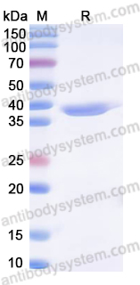
>90% as determined by SDS-PAGE.

Accession

P80108

Form

Lyophilized

Storage buffer

Lyophilized from a solution in PBS pH 7.4, 0.02% SKL, 1mM EDTA, 5% Trehalose, 2% Mannitol.

Reconstitution

Reconstitute in sterile water for a stock solution.

Shipping

In general, proteins are provided as lyophilized powder/frozen liquid. They are shipped out with dry ice/blue ice unless customers require otherwise.

Stability and Storage

Use a manual defrost freezer and avoid repeated freeze thaw cycles. Store at 2 to 8°C for frequent use. Store at -20 to -80°C for twelve months from the date of receipt.

Alternative Names

GPLD1, PIGPLD1, Phosphatidylinositol-glycan-specific phospholipase D, GPI-PLD, GPI-specific phospholipase D, Glycoprotein phospholipase D, PI-G PLD, Glycosyl-phosphatidylinositol-specific phospholipase D

Note

For research use only.

Data Image
  • SDS-PAGE
    SDS PAGE for recombinant Human GPLD1
  • Bioactivity
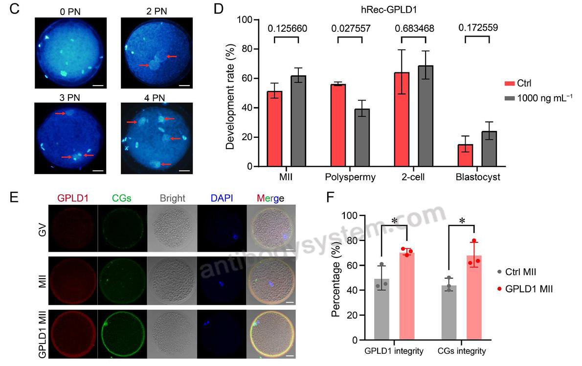
    Figure 4 Role of GPLD1 in regulating polyspermy. C, Immunofluorescence staining of the pronuclei (DAPI) of fertilized embryos, with red arrows indicating the corresponding male and female pronuclei that have undergone depolymerization. Scale bars, 20 μm. D, Bar graph comparing the MII oocyte maturation rate, polyspermy rate, 2-cell cleavage rate, and blastocyst development rate following exogenous GPLD1 supplementation vs. the control group. The numbers represent the P-value of the comparison between the two groups, with a multiple comparison test. E, Immunofluorescence analysis of GPLD1 and CGs across various developmental stages and under different treatment conditions. Scale bars, 20 μm. F, Bar graph comparing the complete distribution rate of GPLD1 in the oocyte plasma membrane and the integrity of CGs plasma membrane expression after GPLD1 supplementation at the MII stage. *, P
    Data sourced from citation (PMID: 42141343)
  • Bioactivity
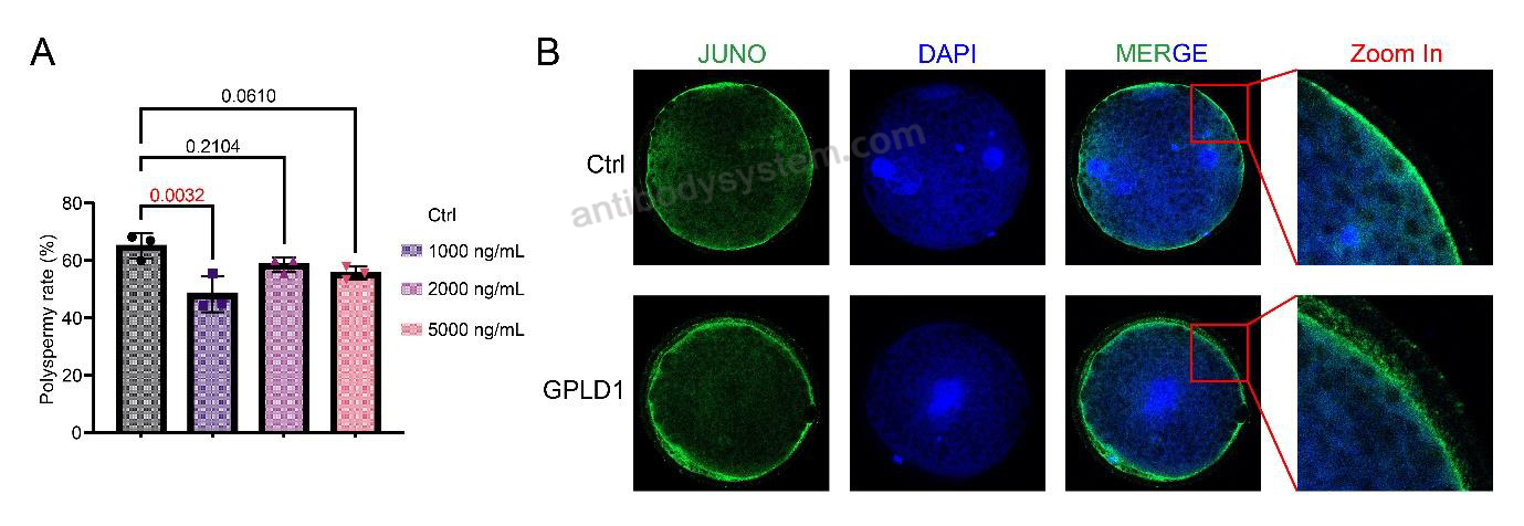
    Supplementary Figure 4. Polyspermy analysis and immunofluorescence staining of JUNO protein in porcine oocytes. (A) The proportion of zygotes exhibiting polyspermy (defined as the presence of more than two pronuclei) was statistically compared across treatment groups. Statistical significance was determined by one-way ANOVA test, and the numbers marked by the horizontal lines represent the P-values. (B) Immunofluorescence staining was performed on porcine zygotes to detect JUNO protein and pronuclei (PN); green fluorescence represents JUNO protein, and blue fluorescence represents DNA in the cell nucleus.
    Data sourced from citation (PMID: 42141343)
References
Datasheet
$ 313
Product specifications
100 μg 313 1 mg 1629

Contact Information

Order: order@antibodysystem.com

Mail: support@antibodysystem.com

Distributor list

For research use only. Not for human or drug use.

Need help with your order?

Find out more about placing an order here

Recombinant Human GPLD1 Protein, N-His [YHF70501]
Terms of sale Website terms of use Cookie policy Privacy
Copyright © 2025 AntibodySystem SAS. All Rights Reserved.            All Products are for Research Use Only