
Please ensure Javascript is enabled for purposes of website accessibility
Home / Products / Antibody / Recombinant Antibody

Anti-Human PTHrP/PTHLH (1-34) Antibody (SAA0326)

Catalog #:   RHC93101 Specific References (0) DATASHEET
Host species: Mouse
Isotype: IgG1
Applications: Neutralization, WB
Accession: P12272
Overview

Catalog No.

RHC93101

Species reactivity

Human, Mouse

Host species

Mouse

Isotype

IgG1

Clonality

Monoclonal

Applications

Neutralization, WB

Target

Parathyroid hormone-like protein, PTHrP, PTH-rP, Parathyroid hormone-related protein, PTHrP[107-139], PLP, PTHRP, PTHLH

Concentration

2.53 mg/ml

Endotoxin level

Please contact with the lab for this information.

Purity

>95% as determined by SDS-PAGE.

Purification

Protein A/G purified from cell culture supernatant.

Accession

P12272

Form

Liquid

Storage buffer

0.01M PBS, pH 7.4.

Stability and Storage

Use a manual defrost freezer and avoid repeated freeze-thaw cycles. Store at 4°C short term (1-2 weeks). Store at -20°C 12 months. Store at -80°C long term.

Clone ID

SAA0326

Note

For research use only.

Data Image
  • Bioactivity
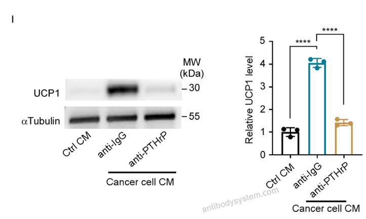
    (I) Adipocytes were treated with control CM or CM from bladder cancer cells in the presence or absence of neutralizing anti-PTHrP or control IgG antibody. Representative immunoblots (left) and quantification (right) of UCP1 in indicated adipocytes.
    Data sourced from citation (PMID: 41690005)
  • Bioactivity
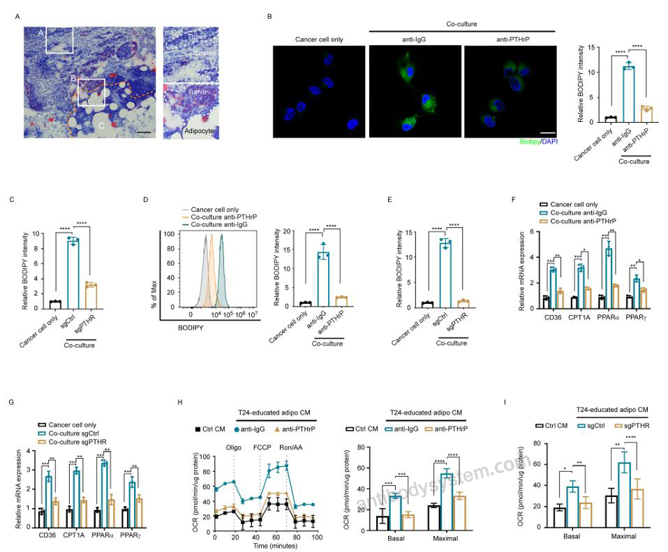
    FFA released from beige adipocytes was taken up by bladder cancer cells and upregulated lipid metabolism. (A) Representative Oil Red O staining image of frozen sections from individuals with bladder cancer. Regions of interest are labeled as follows: A, tumor area distant from adipocytes; B, tumor area adjacent to adipocytes; C, adipocyte-rich area. Yellow dashed lines, the tumor-adipocyte interface; white dashed lines, tumor regions; white square, related magnifications. Scale bar, 100 μm. (B-E) Bladder cancer cells were co-cultured with or without BODIPY-labeled adipocytes, either (B, D) with/without neutralizing anti-PTHrP antibody treatment or (C, E) with/without PTHR knockout in adipocytes. Relative BODIPY fluorescence intensity in bladder cancer cells was quantified via immunofluorescence staining (B, C) or flow cytometry (D, E), with representative images or histograms (left) and quantification (right) of BODIPY staining presented. Scale bar, 20 μm. (F and G) In the presence or absence of neutralizing anti-PTHrP antibody (F), bladder cancer cells were co-cultured with or without PTHR-knockout or wild-type adipocytes (G). The mRNA levels of CD36, CPT1A, PPARɑ, and PPARγ in indicated bladder cancer cells were evaluated by qRT-PCR. (H and I) In the presence or absence of anti-PTHrP antibody (H), bladder cancer cells were co-cultured for 48 h with or without PTHR-knockout or wild-type adipocytes (I). CM from T24-educated adipocytes were then collected to treat T24 cells for 24 h. Representative oxygen consumption curves (left) and quantification of basal OCR (middle) and maximal OCR (right) of indicated bladder cancer cells. Oligo, oligomycin; FCCP, fluorocarbonyl cyanide phenylhydrazone; Rot/AA, rotenone and antimycin A. Data were expressed as means ± SEM (B-I). *p < 0.05, **p < 0.01, ***p < 0.001, ****p < 0.0001 by one-way ANOVA (B-I).
    Data sourced from citation (PMID: 41690005)
  • Bioactivity
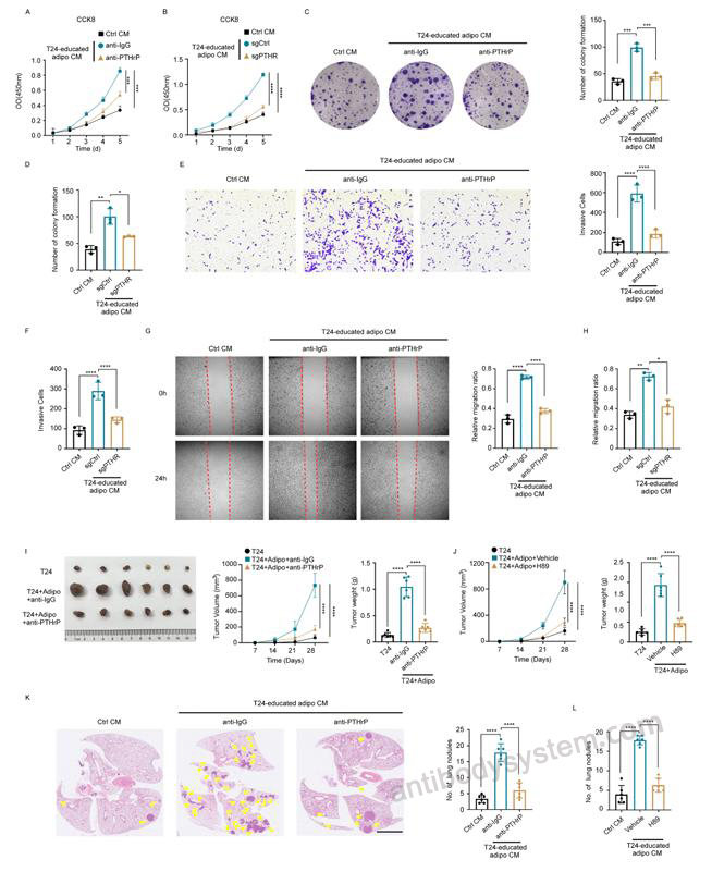
    Inhibition of adipocyte browning suppressed bladder cancer progression. (A-H) In the presence or absence of anti-PTHrP antibody (A, C, E, and G), bladder cancer cells were co-cultured with or without PTHR-knockout or wild-type adipocytes (B, D, F, and H). CM from T24-educated adipocytes were then collected to treat T24 cells. (A and B) The proliferation rates of T24 cells were evaluated by CCK8 assay. (C and D) The proliferation ability of T24 cells was detected by colony formation assay. Representative images and quantification of bladder cancer cells were shown. (E and F) Representative images and quantification of the number of invading T24 cells were clarified by Transwell assay. (G and H) Representative images and quantification of T24 cell migration ability were determined via scratch assay. (I and J) Nude mice were subcutaneously injected with T24 cells alone or co-injected with adipocytes, with or without anti-PTHrP antibody (I) or H89 (J). (I and J) Representative tumor images (left), tumor volume quantification (middle), and tumor weight quantification (right). (K and L) T24 cells were treated with CM from T24-educated adipocytes with or without anti-PTHrP antibody (K) or H89 (L) for two weeks. Nude mice were intravenously injected with indicated bladder cancer cells. Representative H&E-stained images of lung metastases and quantification of lung metastasis lesions are shown. Scale bar, 2.5 mm. Data were expressed as means ± SEM (A-L). *p < 0.05, **p < 0.01, ***p < 0.001, ****p < 0.0001 by one-way ANOVA (A-L).
    Data sourced from citation (PMID: 41690005)
References
Datasheet
$ 372
Product specifications
100 μg 372 1 mg 1860

Contact Information

Order: order@antibodysystem.com

Mail: support@antibodysystem.com

Distributor list

For research use only. Not for human or drug use.

Need help with your order?

Find out more about placing an order here

Anti-Human PTHrP/PTHLH (1-34) Antibody (SAA0326) [RHC93101]
Terms of sale Website terms of use Cookie policy Privacy
Copyright © 2025 AntibodySystem SAS. All Rights Reserved.            All Products are for Research Use Only