
Please ensure Javascript is enabled for purposes of website accessibility
Home / Products / Antibody / Recombinant Antibody

Anti-Human Fibrin Antibody (59D8)

Catalog #:   RHB99201 Specific References (0) DATASHEET
Host species: Mouse
Isotype: IgG1, kappa
Applications: IF, IHC, RIA, WB
Accession: P02675
Overview

Catalog No.

RHB99201

Species reactivity

Human

Host species

Mouse

Isotype

IgG1, kappa

Clonality

Monoclonal

Applications

IF, IHC, RIA, WB

Target

FGB, Fibrinogen beta chain, Fibrin

Concentration

3.02 mg/ml

Endotoxin level

Please contact with the lab for this information.

Purity

>95% as determined by SDS-PAGE.

Purification

Protein A/G purified from cell culture supernatant.

Accession

P02675

Form

Liquid

Storage buffer

0.01M PBS, pH 7.4.

Stability and Storage

Use a manual defrost freezer and avoid repeated freeze-thaw cycles. Store at 4°C short term (1-2 weeks). Store at -20°C 12 months. Store at -80°C long term.

Clone ID

59D8

Note

For research use only.

Data Image
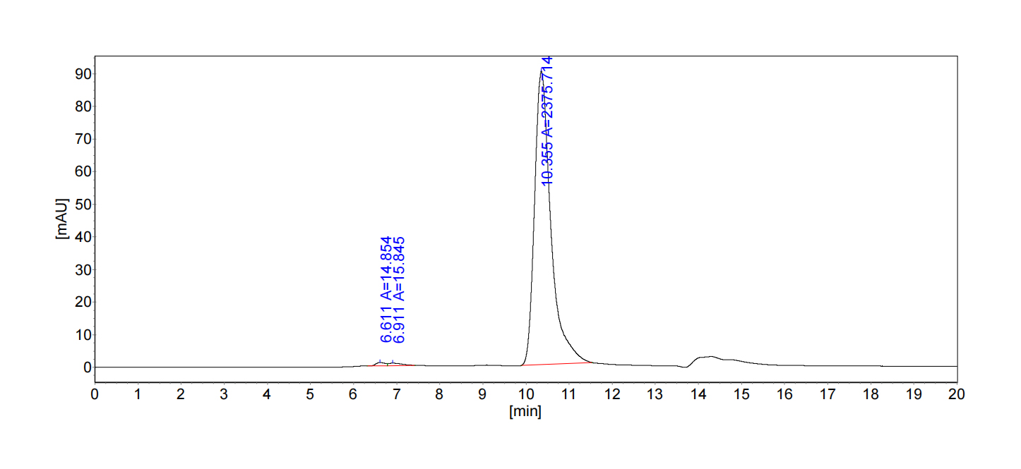
  • SEC-HPLC
    The purity of this product is >95% as determined by SEC-HPLC.
  • Immunofluorescence
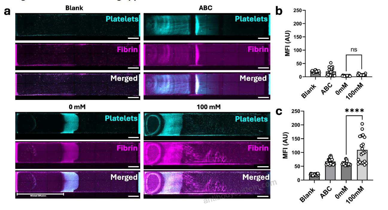
    Wicked distance is viscosity dependent based on platelet-fibrin interactions. a Maximum projection confocal images of the entire RAT test strips (scale bar = 2000 μm) blank (top left), antibody characterisation (ABC, top right), 0 mM (bottom left) and 100 mM (bottom right) CaCl2 recalcified whole blood sample. Platelets (cyan, first row), fibrin (magenta, second row) and merged (last row). Bar charts representing the mean fluorescent intensity for 8-bit images sampled at random ROI across the entire RAT test strip b Platelets and c Fibrin (n = 4, ns = not significant; *p < 0.05; **p < 0.01; ***p < 0.001; ****p < 0.0001, assessed by one-way ANOVA.
    Data sourced from citation (PMID: DOI: 10.1007/s44258-024-00025-3)
  • Immunofluorescence
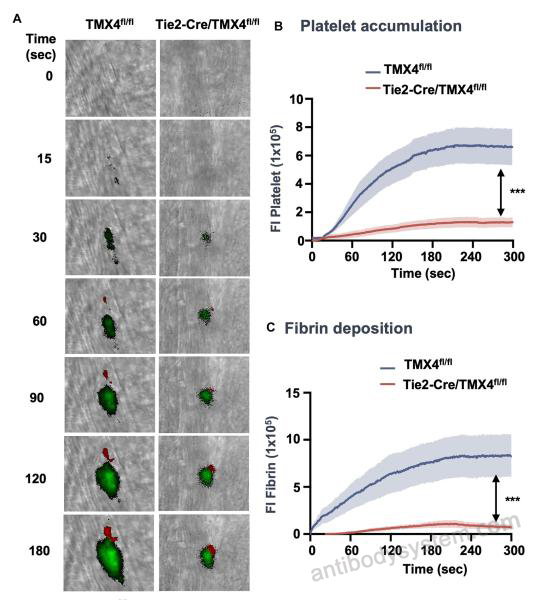
    Tie2-Cre/TMX4fl/fl mice exhibit a decrease in platelet accumulation and fibrin deposition. (A) Platelets and fibrin were detected using Alexa Fluor 488-conjugated anti-CD41 F(ab)2 antibody (green) and Alexa Fluor 647-conjugated anti-fibrin antibody (red), respectively. Representative fluorescence images from intravital microscopy show platelet accumulation (green) and fibrin deposition (red). Original magnification x64. Scale bars, 30 μm. (B, C) The mean ± SEM of fluorescence intensities (FIs) of anti-CD41 antibody (B) and anti-fibrin antibody(C) with the area under the curve were analyzed using a Mann-Whitney rank-sum test. ***P < .001. The data were obtained using 30 thrombi from 3 mice for each experimental condition.
    Data sourced from citation (PMID: 39307246)
  • Immunofluorescence
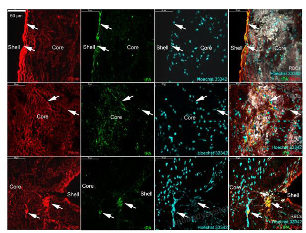
    Figure 2. Localization of tissue-type plasminogen activator in acute ischemic stroke thrombi. A. Representative images of immunofluorescent localization of tissue-type plasminogen activator (tPA, green) in thrombi from IS patients. Top panels: accumulation of tPA in association with compact fibrin (red) in the thrombus outer shell (arrows). Middle panels: tPA bound to fibrin fibers in a red blood cell-rich area of the thrombus core and with compact fibrin (arrows). Lower panels: the white arrows indicate tPA associated with clusters of dense fibrin entangled with extracellular DNA (cyan) close to the thrombus surface. The images shown were acquired in a thrombus from a patient having received intravenous thrombolysis prior to EVT and are representative of 10 different thrombi.
    Data sourced from citation (DOI: DOI: https://doi.org/10.1101/2025.03.28.646062)
  • Bioactivity
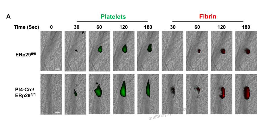
    ERp29 deficiency potentiates platelet accumulation and fibrin deposition and venous thrombosis. (A) Representative images of platelet accumulation (Green) and fibrin generation (Red), as visualized by Dylight-488-conjugated anti-CD42c and Alexa-647-conjugated fibrin (59D8) antibody, respectively, at indicated time points following laser injury in the cremaster arterioles in ERp29fl/fl and Pf4-Cre/ERp29fl/fl mice. Scale bar, 30 μm. The median fluorescence intensity of platelets
    Data sourced from citation (PMID: 40336049)
  • Immunofluorescence
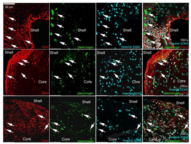
    Figure 3 Localization of plasminogen in acute ischemic stroke thrombi. Representative images of immunofluorescent localization of plasminogen (green) in various thrombi from acute ischemic stroke patients showing the association of plasminogen with dense fibrin (red), nucleated cells and NETs (cyan, arrows).
    Data sourced from citation (PMID: DOI: https://doi.org/10.1101/2025.03.28.646062)
  • Immunofluorescence
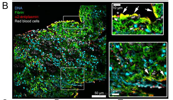
    B. Representative images of α2-antiplasmin (red) localization relative to fibrin (green, arrows) and NETs (cyan, asterisks). Higher magnification views of the squared areas are shown in the insets.
    Data sourced from citation (PMID: DOI: https://doi.org/10.1101/2025.03.28.646062)
References
Datasheet
$ 372
Product specifications
100 μg 372 1 mg 1860

Contact Information

Order: order@antibodysystem.com

Mail: support@antibodysystem.com

Distributor list

For research use only. Not for human or drug use.

Need help with your order?

Find out more about placing an order here

Anti-Human Fibrin Antibody (59D8) [RHB99201]
Terms of sale Website terms of use Cookie policy Privacy
Copyright © 2025 AntibodySystem SAS. All Rights Reserved.            All Products are for Research Use Only