
Please ensure Javascript is enabled for purposes of website accessibility
Home / Products / Antibody / Polyclonal Antibody

Anti-Nipah virus/HeV F/Fusion glycoprotein F0 Polyclonal Antibody

Catalog #:   PVV08101 Specific References (0) DATASHEET
Host species: Rabbit
Isotype: IgG
Applications: ELISA, IHC, WB
Overview

Catalog No.

PVV08101

Species reactivity

Nipah virus(HeV)

Host species

Rabbit

Isotype

IgG

Clonality

Polyclonal

Immunogen

E. coli - derived recombinant Nipah virus/HeV F/Fusion glycoprotein F0 (Gly131-Thr546).

Applications

ELISA, IHC, WB

Recommended Dilution

Application Dilution
ELISA 1:4000-1:8000, IHC: 1:50-1:200, WB: 1:1000-1:4000

Target

Fusion glycoprotein F0, Protein F, Fusion glycoprotein F2, Fusion glycoprotein F1, F

Concentration

1.47 mg/ml

Purification

Purified by antigen affinity column.

Form

Liquid

Storage buffer

0.01M PBS, pH 7.4, 50% Glycerol, 0.05% Proclin 300.

Stability and Storage

Use a manual defrost freezer and avoid repeated freeze thaw cycles. Store at 2 to 8°C for frequent use. Store at -20 to -80°C for twelve months from the date of receipt.

Note

For research use only.

Data Image
  • Western Blot
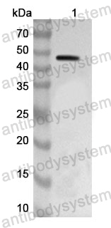
    Recombinant Protein were subjected to SDS PAGE followed by western blot with Nipah_F antibody (PVV08101) at 1μg/ml.

    Lane 1: Recombinant Nipah virus/HeV F/Fusion glycoprotein F0 Protein(YVV08102)

    Second Ab: Goat Anti-Rabbit IgG H&L Polyclonal antibody, HRP (PTB96431) at 0.1 μg/mL.

    Predict MW: 44 kDa
    Observed MW: 44 kDa
  • Western Blot
    Recombinant Protein lysates were subjected to SDS PAGE followed by western blot with F/Fusion glycoprotein F0 antibody (PVV08101) at 1 µg/ml.

    Lane 1: Recombinant Protein

    Second Ab: Goat Anti-Rabbit IgG H&L Polyclonal antibody, HRP (PTB96431) at 0.1 μg/mL.

    Predict MW: 48 kDa
    Observed MW: 48 kDa
  • Western Blot
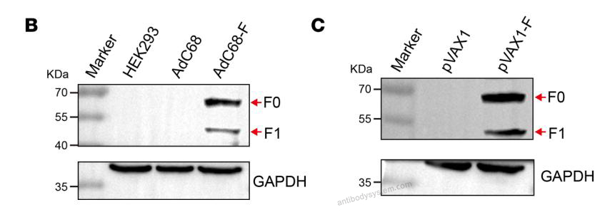
    The specific bands of F protein were detected by Western blotting of AdC68-F-infected HEK293 cells (B) and pVAX1-F-transfected HEK293T cells (C). Both F0 (61 kDa) and F1 (49 kDa) could be detected in AdC68-F-infected and pVAX1-F-transfected cells. The F antigens were detected using a polyclonal rabbit anti-NiV glycoprotein F0 antibody (AntibodySystem, PVV08101; diluted 1:5000).(PMID: 37917215)
  • Western Blot
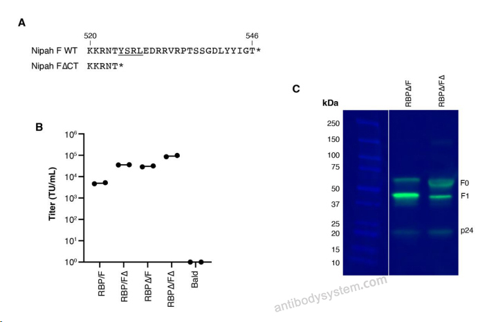
    Fig. S2. F cytoplasmic tail truncation affects pseudovirus titers and cleavage A) Amino-acid alignment of wildtype (WT) and 22 amino-acid cytoplasmic tail deletion (∆CT) at the C-terminus of Nipah virus F. Endocytosis motif 'YSRL' is underlined. B) Pseudovirus titers in CHO-bEFNB3cells of different combinations of RBP and F constructs with or without the cytoplasmic tail deletion. C) Reduced SDS-PAGE western blot on F and p24 in pseudoviruses (see Methods for details on staining). F0 and F1 are uncleaved and cleaved products, respectively. Gel is representative of two experiments
    Data sourced from citation (PMID: 41650235)
  • Western Blot
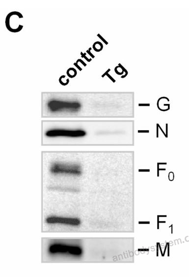
    (C) Cell lysates were collected 24 h p.i. and subjected to western blot analysis. NiV N, F, G, and M were stained with respective specific antibodies, biotinylated secondary antibodies, and HRP-conjugated streptavidin. Chemiluminescent signals were recorded with a Chemidoc system.
    Data sourced from citation (PMID: 41378893)
References
Datasheet
$ 170
Product specifications
50 μg 170 100 μg 280

Contact Information

Order: order@antibodysystem.com

Mail: support@antibodysystem.com

Distributor list

For research use only. Not for human or drug use.

Need help with your order?

Find out more about placing an order here

Anti-Nipah virus/HeV F/Fusion glycoprotein F0 Polyclonal Antibody [PVV08101]
Terms of sale Website terms of use Cookie policy Privacy
Copyright © 2025 AntibodySystem SAS. All Rights Reserved.            All Products are for Research Use Only