
Please ensure Javascript is enabled for purposes of website accessibility
Home / Products / Antibody / Polyclonal Antibody

Anti-DHODH Polyclonal Antibody

Catalog #:   PHF91801 Specific References (0) DATASHEET
Host species: Rabbit
Isotype: IgG
Applications: ELISA, IHC, WB
Accession: Q02127
Overview

Catalog No.

PHF91801

Species reactivity

Human, Mouse, Rat

Host species

Rabbit

Isotype

IgG

Clonality

Polyclonal

Immunogen

E. coli - derived recombinant Human DHODH (Thr31-Arg395).

Applications

ELISA, IHC, WB

Recommended Dilution

Application Dilution
ELISA 1:4000-1:8000, IHC: 1:50-1:100, WB: 1:1000-1:4000

Target

Dihydroorotate oxidase,Dihydroorotate dehydrogenase (quinone),mitochondrial,DHODH,DHOdehase

Purification

Purified by antigen affinity column.

Accession

Q02127

Form

Liquid

Storage buffer

0.01M PBS, pH 7.4, 50% Glycerol, 0.05% Proclin 300.

Stability and Storage

Use a manual defrost freezer and avoid repeated freeze thaw cycles. Store at 2 to 8°C for frequent use. Store at -20 to -80°C for twelve months from the date of receipt.

Note

For research use only.

Data Image
  • WESTERN BLOT
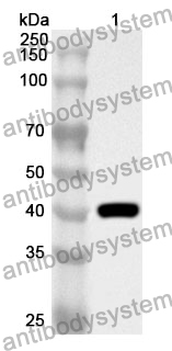
    Recombinant Protein lysates were subjected to SDS PAGE followed by western blot with DHODH antibody (PHF91801) at 1 μg/ml.

    Lane 1: Recombinant protein

    Second Ab: Goat Anti-Rabbit IgG H&L Polyclonal antibody, HRP (PTB96431) at 0.1 μg/mL.

    Predict MW: 42 kDa
    Observed MW: 42 kDa
  • Western Blot
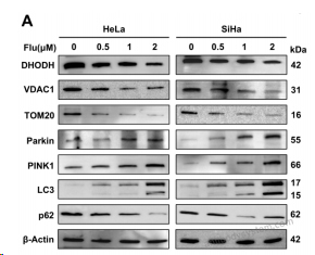
    Fig 3 Flubendazole downregulates DHODH expression and induces mitophagy in cervical carcinoma cells. (A) Western blot analysis of DHODH, VDAC1, TOM20, Parkin, PINK1, LC3, and p62 protein expression levels in HeLa and SiHa cells treated with escalating concentrations (0, 0.5, 1, 2 µM) flubendazole for 48 hours.
    Data sourced from citation (PMID: 40885318)
  • Western Blot
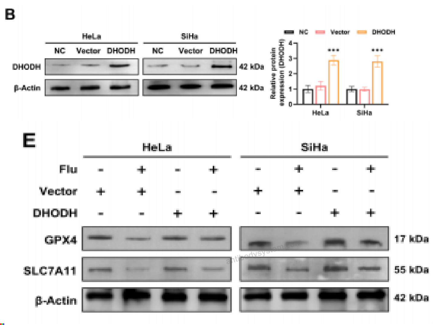
    Fig 4 DHODH overexpression diminishes flubendazole-induced ferroptosis and its antitumor effects on cervical carcinoma cells. western blotting was conducted to detect the expression levels of DHODH (B). (E) Western blot analysis of the expression levels of GPX4 and SLC7A11 in HeLa and SiHa cells overexpressing DHODH alone or co-treated with flubendazole (2 μM, 48 h)
    Data sourced from citation (PMID: 40885318)
  • Western Blot
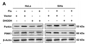
    Fig 5 DHODH overexpression reduces flubendazole-induced mitophagy in cervical carcinoma cells. (A) Western blot analysis of the expression levels of Parkin and PINK1 proteins in HeLa and SiHa cells overexpressing DHODH, with or without co-treatment of 2 μM flubendazole for 48 hours.
    Data sourced from citation (PMID: 40885318)
  • Western Blot
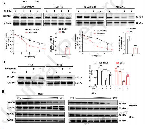
    Fig 6 Flubendazole targets DHODH to promote its protein degradation in cervical carcinoma cells. (C) HeLa and SiHa cells were pretreated with or without flubendazole (2 μM, 24 h) and then co-treated with 50 μg/ml cycloheximide (CHX) for the indicated time (0, 1, 2, 4 h) and observed the degradation rate of DHODH protein. (D) The lysates extracted from HeLa and SiHa cells were incubated with or without 20 μM flubendazole for 1 hour at room temperature, then were subjected to DARTS assay using Pronase E and detected by western blotting. (E) HeLa and SiHa cell lysates (incubated with or without 20 μM flubendazole) were analyzed by CETSA-WB to further validate the interaction between flubendazole and DHODH.
    Data sourced from citation (PMID: 40885318)
  • Western Blot
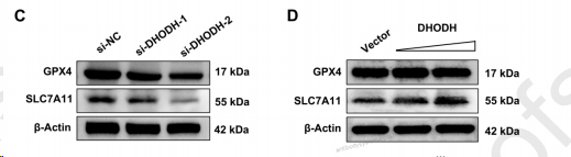
    Fig 8 The regulation of GPX4/SLC7A11 by DHODH. (C) Western blot analysis of GPX4 and SLC7A11 expression in DHODH-knockdown cells or control cells. (D) HeLa and SiHa cells were transfected with 2.5 μg Vector or 2.5 μg DHODH plasmid or 5 μg DHODH plasmid. 48 hours later, cells were lysed and subjected to Western blot to analyze the GPX4 and SLC7A11 expression. Protein levels of GPX4 and SLC7A11 were normalized to β-actin and quantified in the lower panels.
    Data sourced from citation (PMID: 40885318)
  • Immunohistochemistry
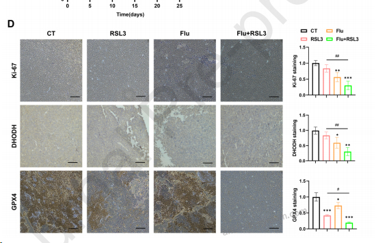
    Fig 9 Enhanced antitumor effects of flubendazole combined with RSL3 in cervical cancer xenograft models. (D) The expressions of Ki-67, DHODH, and GPX4 staining in tumor tissues from the different groups were determined by immunohistochemical staining, and the staining score was calculated.
    Data sourced from citation (PMID: 40885318)
References
Datasheet
$ 170
Product specifications
50 μg 170 100 μg 280

Contact Information

Order: order@antibodysystem.com

Mail: support@antibodysystem.com

Distributor list

For research use only. Not for human or drug use.

Need help with your order?

Find out more about placing an order here

Anti-DHODH Polyclonal Antibody [PHF91801]
Terms of sale Website terms of use Cookie policy Privacy
Copyright © 2025 AntibodySystem SAS. All Rights Reserved.            All Products are for Research Use Only