
2026 Nobel Prize in Physiology or Medicine: How Optogenetics Uses Light to Control Neuronal Activity
The 2026 Nobel Prize in Physiology or Medicine has been awarded to Karl Deisseroth, Peter Hegemann, and Georg Nagel "for their discoveries concerning light-gated ion channels and optogenetics."

The work recognized by this year's Nobel Prize traces back to studies of how the green alga Chlamydomonas reinhardtii senses light. Research by Peter Hegemann and Georg Nagel helped establish Channelrhodopsins as light-gated ion channels, providing the molecular basis for optogenetics. Karl Deisseroth and his collaborators subsequently demonstrated how these light-sensitive proteins could be used to control neuronal activity with millisecond-scale precision.
Together, these discoveries established a new experimental approach for neuroscience: combining genetic targeting with optical control to manipulate defined populations of neurons and investigate their roles in neural circuits and behavior.

From Channelrhodopsin to Optogenetics
The foundation of optogenetics lies in a basic question in cell biology: how can a cell detect light and convert that signal into a physiological response?
In the 1990s, Peter Hegemann investigated phototaxis in Chlamydomonas reinhardtii, a unicellular green alga that changes its swimming direction in response to light. His work helped identify the molecular machinery responsible for light sensing.
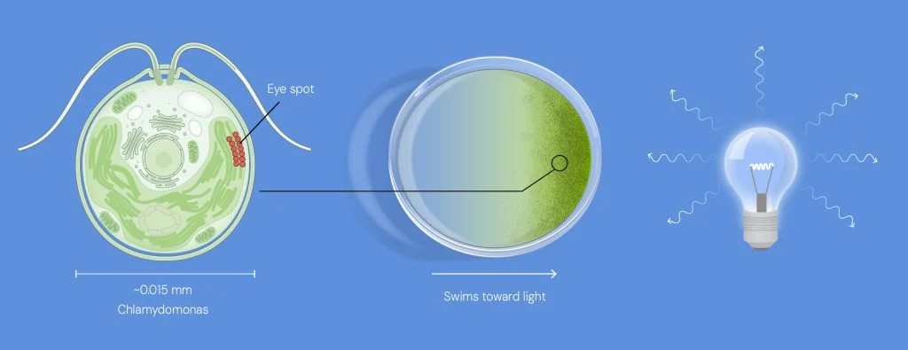
The alga Chlamydomonas senses light using its eyespot. When light is sufficiently strong, it swims towards the source. © The Nobel Committee for Physiology or Medicine. Ill. Mattias Karlén
Subsequent studies by Georg Nagel, Hegemann, and their collaborators characterized microbial rhodopsins from Chlamydomonas. Their work established that Channelrhodopsins are not simply light-sensitive proteins. They can function as light-gated ion channels, directly coupling light stimulation to ion movement across the cell membrane.
This property was particularly important because it provided a direct route from an optical signal to membrane electrical activity.
In the case of Channelrhodopsin-2 (ChR2), light activation opens the channel and allows cations to cross the membrane. The resulting change in membrane potential can alter the electrical state of the cell.
The basic mechanism can therefore be summarized as:
Light → Channelrhodopsin activation → Ion flow → Change in membrane potential
For neuroscience, this raised an important possibility: could a light-gated ion channel from algae be used to control the electrical activity of neurons?
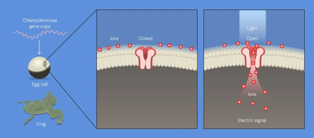
When Nagel and Hegemann introduced the Chlamydomonas genes in frog eggs, they began to produce ion channel proteins that ended up on the surface of the eggs. When the researchers illuminated the ion channels, they opened. Ions then flowed through the channels, creating an electric signal. © The Nobel Committee for Physiology or Medicine. Ill. Mattias Karlén
How Light Became a Tool for Controlling Neurons
Karl Deisseroth and his collaborators helped translate the properties of microbial opsins into a practical tool for neuroscience.
The key concept was to combine two types of control.
First, genetic approaches can be used to restrict opsin expression to a defined neuronal population. Second, light can be delivered to those cells to control their activity at a specific time.
In 2005, Edward Boyden, Feng Zhang, Ernst Bamberg, Georg Nagel, Karl Deisseroth, and colleagues reported a landmark study in Nature Neuroscience. Using ChR2 and gene delivery, they demonstrated optical control of mammalian neuronal activity on a millisecond timescale. The study showed that light stimulation could control neuronal spiking as well as excitatory and inhibitory synaptic transmission.
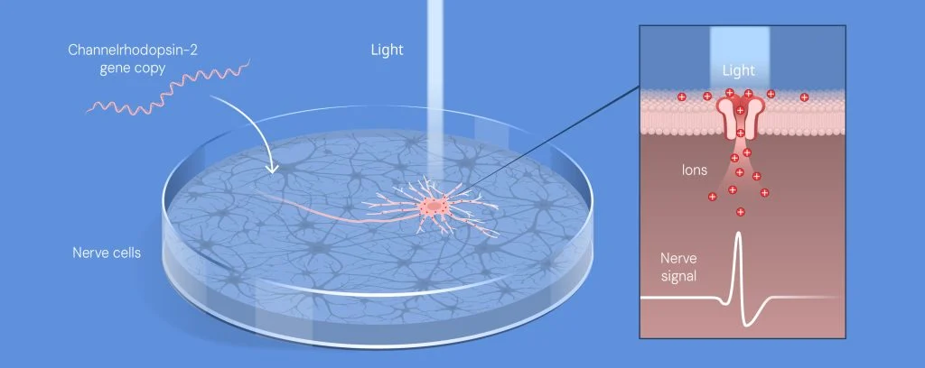
Deisseroth introduced the gene encoding channelrhodopsin-2 into nerve cells that he cultured in a petri dish, making the nerve cells sensitive to light. Using blue light, the researchers were able to trigger a rapid nerve signal. © The Nobel Committee for Physiology or Medicine. Ill. Mattias Karlén
This work established a core principle of optogenetics:
Genetic targeting determines which cells can be manipulated; optical stimulation determines when they are manipulated.
The combination provides both cell-type specificity and high temporal precision, two properties that are particularly valuable for studying dynamic neural circuits.
From Correlation to Causality
Before optogenetics, neuroscience had already developed powerful methods for recording neuronal activity. Researchers could identify neuronal populations that became active during memory, movement, reward, sleep, or other behaviors. However, observing activity does not necessarily establish its functional role.
If a population of neurons becomes active during a particular behavior, are those neurons actually driving the behavior, or are they responding to another process occurring at the same time?
Optogenetics provides a way to address this question experimentally.
Researchers can selectively express an opsin in a defined neuronal population and then manipulate those cells while monitoring neural activity, circuit dynamics, or behavior.
The experimental framework shifts from:
Observe neuronal activity → measure behavior
to:
Select a neuronal population → manipulate its activity → measure the resulting response
This ability to perturb specific components of a neural circuit has made optogenetics an important tool for investigating causal relationships in neuroscience.
It has been applied to questions involving memory formation, reward and addiction, motor control, sleep and arousal, and other aspects of brain function. The approach has also contributed to experimental studies of neurological and psychiatric disorders by allowing researchers to investigate how specific neuronal circuits contribute to disease-associated phenotypes.
Why Optogenetics Matters for Neuroscience
The significance of optogenetics extends beyond the ability to activate or inhibit neurons with light.
Its broader contribution is methodological: it provides researchers with a way to connect molecular tools, defined cell populations, neural circuits, and behavior within the same experimental framework.
The development of increasingly diverse microbial opsins, together with advances in genetic targeting, viral vectors, optical delivery, and circuit-level recording, has continued to expand the range of questions that can be addressed using optogenetic approaches.
The scientific progression recognized by the 2026 Nobel Prize can be summarized as:
The significance of this progression lies in how a fundamental discovery in microbial photobiology became a versatile research tool for neuroscience.
From a Light-Sensing Protein to a Neuroscience Research Tool
The work of Hegemann, Nagel, and Deisseroth illustrates how fundamental biological research can lead to technologies that reshape experimental approaches in another field.
A protein involved in light sensing in a unicellular alga became a molecular tool for controlling neuronal activity. By combining light-gated ion channels with genetic targeting and optical stimulation, researchers gained a new way to test how specific neurons and neural circuits contribute to brain function and behavior.
This transition—from observing neuronal activity to experimentally manipulating it—is central to the impact of optogenetics and to the scientific work recognized by the 2026 Nobel Prize in Physiology or Medicine.
From Neural Circuits to Disease Mechanisms
As neuroscience moves from neural circuits toward molecular mechanisms, combining precise neuronal manipulation with molecular characterization may provide new ways to investigate disease mechanisms. In neurodegenerative disease research, targets such as Aβ, Tau/p-Tau, APOE, and TREM2 provide molecular readouts relevant to Alzheimer’s disease, while TH, dopamine receptors, and α-synuclein support research into dopaminergic signaling and Parkinson’s disease. Other targets, including TDP-43, GFAP, IBA1, GPR17, and NfL, can help characterize molecular changes associated with ALS/FTD, neuroinflammation, and neuronal injury.
AntibodySystem provides antibodies and related research reagents targeting these and other neuroscience-associated proteins, offering molecular research tools that can complement circuit-level approaches such as optogenetics.
AntibodySystem Research Tools for Neuroscience
| Research Area | Target | Catalog No. | Product Name |
|---|---|---|---|
| Alzheimer’s disease | Aβ | RHC12520 | Anti-β-Amyloid (1-16aa) Antibody (6E10#) |
| Tau / p-Tau | RHC82412 | Anti-Human Phospho-Tau (pS202/pT205) Antibody (AT8) | |
| APOE | RHB98601 | Anti-Human APOE Antibody (SAA0799) | |
| TREM2 | RHJ69202 | Anti-Human TREM2 Antibody (SAA0340) | |
| Parkinson’s disease / Dopaminergic signaling | TH | RHC22902 | Anti-TH Antibody (R3A02) |
| DRD2 | RHD04802 | Anti-Human DRD2 Nanobody (SAA1361) | |
| α-Synuclein | YHE22001 | Recombinant Human SNCA Protein, N-His | |
| ALS / FTD | TDP-43 | YHG42201 | Recombinant Human TDP43 Protein, N-His |
| CHCHD10 | YHK85501 | Recombinant Human CHCHD10 Protein, N-GST & C-His | |
| Neuroinflammation / CNS injury | GFAP | RGD03101 | Anti-Human GFAP Antibody (SAA0567) |
| IBA1 | PHF07401 | Anti-AIF1/IBA1 Polyclonal Antibody | |
| GPR17 | YHG47702 | Recombinant Human GPR17 Protein, N-GST & C-His | |
| Neuronal injury / Neurodegeneration | NfL / NEFL | RHC23302 | Anti-NEFL Antibody (R3A03) |
| RBPMS | PHK72701 | Anti-Human RBPMS Polyclonal Antibody |
Reference
1. Nagel G, et al. Channelrhodopsin-1: A Light-Gated Proton Channel in Green Algae. Science. 2002;296:2395-2398.
2. Nagel G, et al. Channelrhodopsin-2: A Light-Gated Proton Channel in Green Algae. Proc Natl Acad Sci USA. 2003;100:13940-13945.
3. Boyden ES, Zhang F, Bamberg E, Nagel G, Deisseroth K. Millisecond-timescale, genetically targeted optical control of neural activity. Nature Neuroscience. 2005;8:1263-1268. DOI: 10.1038/nn1525.
4. Kato HE, Zhang F, Yizhar O, et al. Crystal structure of the channelrhodopsin light-gated cation channel. Nature. 2012;482:369-374. DOI: 10.1038/nature10870.
5. Deisseroth K. Optogenetics: 10 years of microbial opsins in neuroscience. Nature Neuroscience. 2015;18:1213-1225. DOI: 10.1038/nn.4091.
6. Nobel Prize. The Nobel Prize in Physiology or Medicine 2026 - Popular information.
7. Naddaf M, Callaway E. Medicine Nobel awarded for brain 'switch' that controls neurons with light. Nature. 2026. DOI: 10.1038/d41586-026-03091-2.


