
Chandipura Virus (CHPV): Structure, Pathogenesis, and Therapeutic Landscape
Chandipura virus (CHPV), a highly neurotropic rhabdovirus, has emerged as a serious public health threat across the Indian subcontinent. A recent outbreak in Gujarat, western India, has underscored this danger. As of August 3, 2026, health officials there had reported 184 suspected cases, including 35 laboratory-confirmed infections, 22 deaths, and 7 patients in critical condition. All affected individuals were under 15 years of age.

CHPV belongs to the family Rhabdoviridae, genus Vesiculovirus. Transmitted primarily by phlebotomine sandflies, it causes rapidly progressive neurological disease with high mortality, establishing it as a major emerging cause of viral encephalitis. No specific antivirals or vaccines are currently approved. Dissecting CHPV protein architecture and pathogenesis, and developing antibody-based diagnostics and therapeutics, are therefore urgent priorities.
Viral Protein Architecture and Molecular Assembly
The CHPV genome encodes five major structural proteins: nucleoprotein (N), phosphoprotein (P), matrix protein (M), glycoprotein (G), and large polymerase (L). Each plays an essential role in the viral life cycle.
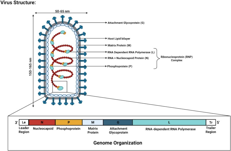
- Nucleoprotein (N): Encapsidates viral RNA to form the nucleocapsid, the core structural component of the viral ribonucleoprotein complex.
- Phosphoprotein (P): Acts as a dedicated chaperone for N, preventing its non-specific aggregation during replication to ensure proper nucleocapsid assembly. It also regulates viral transcription and replication.
- Matrix Protein (M): Lies beneath the envelope and maintains virion structural integrity.
- Glycoprotein (G): The sole spike on the virion surface, mediating receptor recognition and fusion between the viral envelope and the host cell membrane. Computational modeling further reveals that G proteins from Indian and West African strains diverge at the receptor-binding interface, a difference that may explain the enhanced pathogenicity of the Indian isolate in humans.
- Polymerase (L): Catalyzes viral RNA replication and transcription.
Transmission
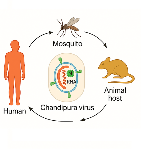
Although ticks and mosquitoes have also been implicated, sandflies remain the principal vectors, spanning the genera Phlebotomus and Sergentomyia. Live virus and viral RNA have been isolated from Sergentomyia species in endemic areas, and experimental studies show that Phlebotomus papatasi can infect mice orally. Human-to-human transmission has not been documented, so vector control is the cornerstone of prevention.
Infection begins with high fever, headache, nausea, and malaise, then deteriorates rapidly into seizures and coma. Without prompt intervention, death often occurs within 48–72 hours of symptom onset. Children under 15 are especially vulnerable because of immature immune and neurological development.
Pathogenic Mechanisms
CHPV is a neurotropic virus with specific affinity for the nervous system.
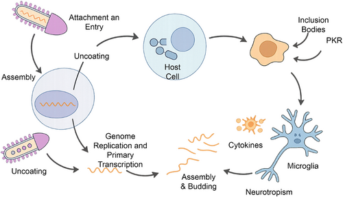
- Step 1: Entry. Following inoculation by sandfly bite, G protein binds to host cell receptors to mediate entry. Recent work identifies LRP1 (low-density lipoprotein receptor-related protein 1) as a likely key receptor for human cell invasion.
- Step 2: Blood-Brain Barrier Penetration. Like other neurotropic viruses, CHPV breaches the blood-brain barrier (BBB) to access the central nervous system.
- Step 3: Neuronal Apoptosis. Within the brain, the virus drives neuronal death through the Fas-mediated extrinsic apoptotic pathway, activating caspase-3 and caspase-8. Neuroinflammation is a hallmark of infection, with prominent involvement of the cerebrum and brainstem.
- Step 4: Cytoplasmic Inclusion Bodies. Infected cells develop cytoplasmic inclusion bodies (IBs) that serve as replication compartments. Viral and host proteins co-localize within these heterogeneous inclusions, enabling the virus to evade antiviral stress granules (SGs).
Current Diagnostic Approaches
CHPV infection is acute and rapidly progressive, making early diagnosis critical.
- Early stage (days 0–4): Real-time RT-PCR for viral RNA is the diagnostic mainstay.
- Mid-to-late stage or surveillance: The IgM ELISA developed by ICMR-NIV is widely used for field monitoring and retrospective confirmation of convalescent cases.
- Research use: Recombinant G protein-based ELISAs are available for antibody detection but remain investigational and lack regulatory approval for clinical diagnosis.
The plaque reduction neutralization test (PRNT) remains the gold standard for neutralizing antibody detection, though it is laborious and slow. The newer microneutralization ELISA (MN-ELISA) offers a faster alternative for serosurveys and vaccine trials.
Clinically, CHPV-induced acute encephalitis syndrome (AES) closely resembles Japanese encephalitis and other etiologies, requiring multiplex molecular panels for differential diagnosis.
Therapeutic and Vaccine Development
With historical case-fatality rates of 50–75% during outbreaks and rapid clinical progression, effective antivirals are urgently needed. None are currently approved, and management remains primarily supportive: controlling intracranial pressure, administering anticonvulsants, maintaining respiratory and hemodynamic stability, and preventing secondary infections.
Vaccine development is still preclinical, driven by reverse vaccinology and multi-epitope peptide design. Immunogenic T- and B-cell epitopes have been mapped across the N, P, and G proteins. Conserved neutralizing epitopes on G are particularly attractive subunit vaccine targets because they mediate viral entry while showing sufficient inter-strain divergence. Molecular docking and dynamics simulations confirm that candidate epitopes bind stably to MHC molecules and display favorable predicted immunogenicity. Novel SCID mouse models now provide robust platforms for in vivo efficacy testing.
The main challenges are optimizing immunogenicity to elicit durable neutralizing antibodies and validating CNS-protective efficacy against neuroinvasion. Accelerating translational research from computational modeling to animal studies is essential to combat this lethal pathogen.
AntibodySystem Product Portfolio
As CHPV research advances, high-quality antibodies and reagents have become essential for detection, mechanistic investigation, and drug development. AntibodySystem offers specialized products to support researchers in protein structural analysis, pathogenesis studies, and diagnostic reagent development.
| Catalog No. | Product Name |
|---|---|
| EVV40701 | Recombinant CHPV G/Glycoprotein Protein, C-His |
| EVV40702 | Recombinant CHPV G/Glycoprotein Protein, C-Fc |
| YVV40701 | Recombinant CHPV G/Glycoprotein Protein, N-His |
| YVV42801 | Recombinant CHPV P/Phosphoprotein Protein, N-His |
| YVV42901 | Recombinant CHPV M/Matrix protein Protein, N-His |
| YVV43001 | Recombinant CHPV N/Nucleoprotein Protein, N-His |
| PVV40701 | Anti-CHPV G/Glycoprotein Polyclonal Antibody |
| PVV42801 | Anti-CHPV P/Phosphoprotein Polyclonal Antibody |
| PVV42901 | Anti-CHPV M/Matrix protein Polyclonal Antibody |
| PVV43001 | Anti-CHPV N/Nucleoprotein Polyclonal Antibody |
Explore all CHPV products at AntibodySystem →
References
- Chandipura Virus: An Emerging Neurological Threat Transmitted by Sandflies in India. PMID: 42447497
- A Multi-Domain Approach for Designing of Novel Epitopes-Based Vaccine Against Chandipura Virus.
- Chandipura virus: A comprehensive review. PMID: 41161670
- Identification of a conserved neutralizing epitope in the G-protein of Chandipura virus.


