
Why Are There No Bands in My Western Blot? A Complete Troubleshooting Guide
Western Blot Troubleshooting · Signal Failure Guide
Western blot (WB) is one of the most widely used techniques for protein detection. However, one of the most frustrating outcomes is completing the entire experiment only to find no detectable bands.
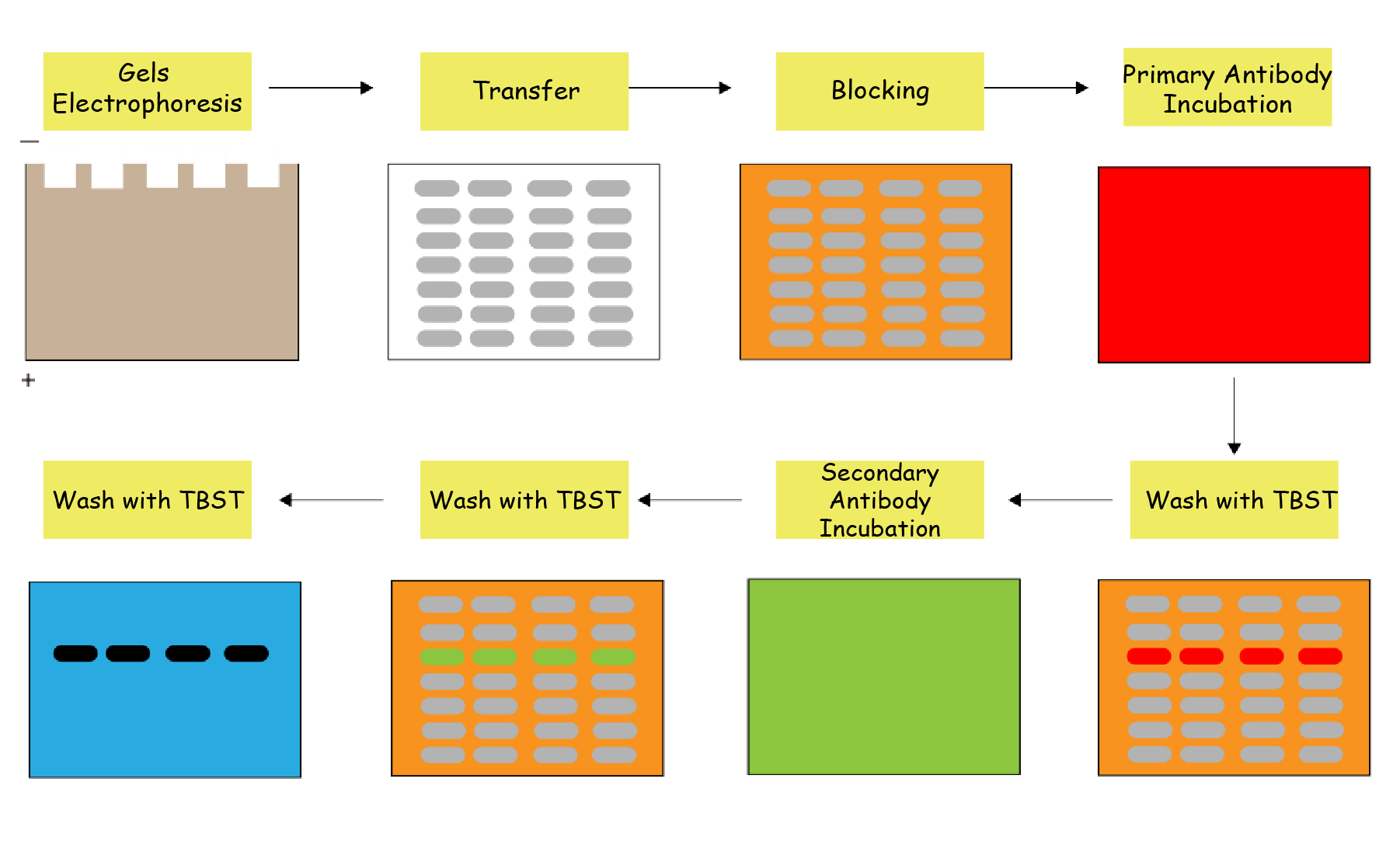
If your membrane shows no signal, don't panic. The problem is usually caused by one or more issues during sample preparation, electrophoresis, transfer, antibody incubation, or detection.
This guide walks through the most common causes and practical solutions.
Problem
After developing the membrane, there are:
- No target bands
- No molecular weight marker (if chemiluminescent marker is used)
- Completely blank membrane
Possible Cause 1: Insufficient Target Protein
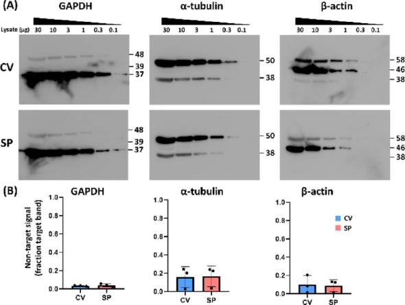
If the target protein is expressed at very low levels, the amount loaded may not be sufficient for detection.
What to check
- Is the protein naturally expressed in your cell or tissue?
- Was protein degradation prevented during extraction?
- Was protein concentration accurately measured?
Solutions
- Increase protein loading amount.
- Use fresh lysates and include protease inhibitors.
- Verify protein concentration using BCA or Bradford assay.
- Consider enriching membrane or nuclear proteins if your target is localized.
Possible Cause 2: Poor Protein Transfer
Even if electrophoresis is successful, proteins may fail to transfer efficiently onto the membrane.
What to check
- Transfer time
- Transfer current or voltage
- Membrane orientation
- Air bubbles between gel and membrane
Solutions
- Confirm the membrane was placed on the correct side of the gel.
- Remove all air bubbles before transfer.
- Increase transfer time for large proteins.
- Reduce methanol concentration when transferring proteins above 150 kDa.
- Use PVDF membranes for better protein binding.
Possible Cause 3: Primary Antibody Problems
Primary antibody issues are among the most common reasons for failed Western blots.
What to check
- Is the antibody validated for WB?
- Was the antibody diluted correctly?
- Has the antibody expired or undergone repeated freeze-thaw cycles?
Solutions
- Use antibodies validated specifically for Western blot.
- Optimize antibody dilution according to the manufacturer's recommendation.
- Store antibodies appropriately and avoid repeated freeze-thaw cycles.
- Include a positive control sample.
Possible Cause 4: Secondary Antibody Issues
Incorrect secondary antibodies or detection reagents can eliminate all signal.
What to check
- Species compatibility
- HRP or fluorescent label
- Antibody dilution
Solutions
- Confirm the secondary antibody matches the host species of the primary antibody.
- Ensure the detection substrate matches the enzyme label.
- Prepare fresh secondary antibody working solution.
Possible Cause 5: Detection Reagent Failure
Sometimes the blot is perfectly fine, but the detection reagent has lost activity.
What to check
- ECL substrate expiration
- Imaging instrument settings
- Exposure time
Solutions
- Use freshly prepared ECL substrate.
- Increase exposure time.
- Verify the imaging system using a positive control.
Possible Cause 6: Incorrect Blocking or Washing
Excessive blocking or over-washing can reduce antibody binding.
Solutions
- Block for 1 hour at room temperature rather than overnight when possible.
- Avoid excessive washing.
- Use appropriate blocking buffers for phosphoproteins or glycoproteins.
Quick Troubleshooting Checklist
| Observation | Most Likely Cause | Recommended Action |
|---|---|---|
| No bands at all | Transfer failure | Check transfer orientation and efficiency |
| Ladder visible but no target | Antibody issue | Verify antibody specificity and dilution |
| Weak bands | Low protein expression | Increase protein loading |
| All samples negative | Detection failure | Replace ECL substrate |
| Positive control negative | Experimental error | Recheck every step of the workflow |
Best Practices to Prevent Blank Western Blots
Successful Western blot experiments rely on optimizing every step of the workflow:
- Use high-quality protein samples.
- Verify protein concentration before loading.
- Include positive and negative controls.
- Optimize antibody concentrations.
- Confirm transfer efficiency before antibody incubation.
- Use validated antibodies and fresh detection reagents.
By systematically checking each step, most "no band" problems can be identified and corrected quickly.
Frequently Asked Question
Q: My housekeeping protein is visible, but my target protein is not. What does this mean?
This usually indicates that protein extraction and transfer were successful. The issue is more likely related to low target expression, antibody specificity, or antibody concentration. Testing a positive control sample and optimizing antibody conditions are recommended.


