
Why Is There No Staining in My Immunohistochemistry (IHC) Experiment?
Immunohistochemistry Troubleshooting · Staining Failure Guide
Immunohistochemistry (IHC) is widely used to visualize protein expression and localization in tissue sections. However, one of the most common challenges researchers encounter is complete absence of staining, even when the target protein is expected to be present.
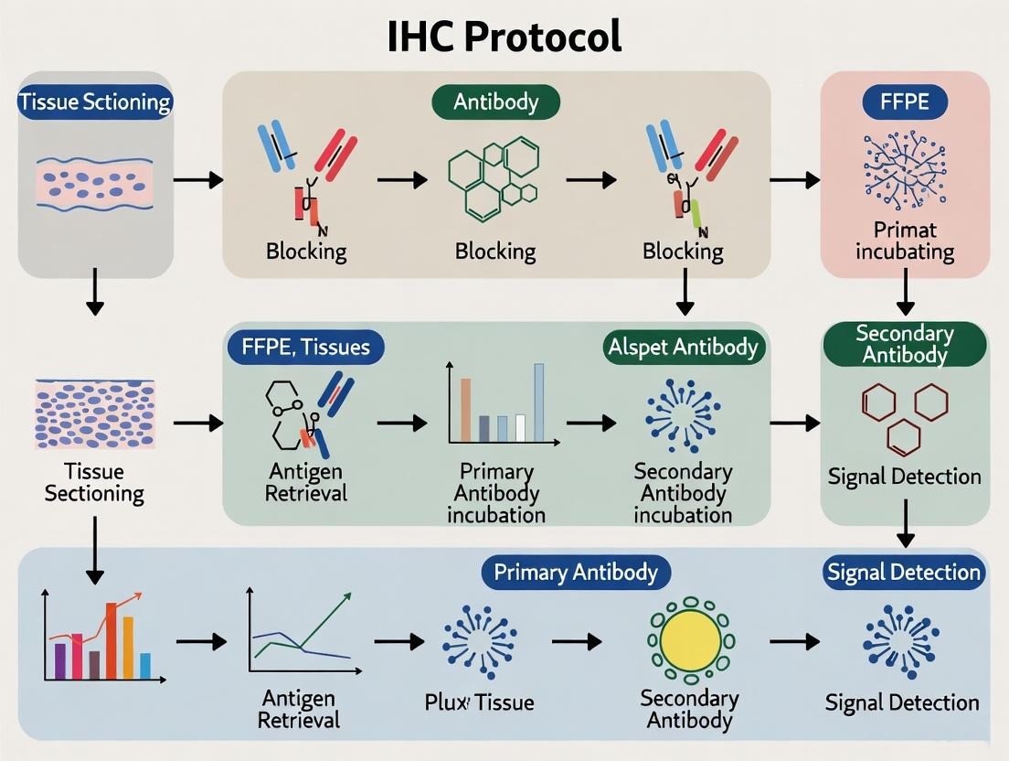
A lack of staining does not necessarily indicate that the target protein is absent. In many cases, the issue arises from tissue processing, antigen retrieval, antibody performance, or detection conditions.
This guide outlines the most common causes of failed IHC staining and provides practical solutions to help you identify the problem.
Problem
After completing the staining procedure:
- No positive staining is observed.
- Both the test sample and positive control are negative.
- Tissue morphology appears normal, but no specific signal is detected.
Possible Cause 1: Antigen Retrieval Was Insufficient
Formalin fixation creates protein cross-links that can mask antigen epitopes. Without effective antigen retrieval, antibodies may not be able to access their target.
What to check
- Was antigen retrieval performed?
- Was the retrieval buffer appropriate for the target antigen?
- Were the temperature and retrieval time sufficient?
Solutions
- Optimize heat-induced epitope retrieval (HIER) conditions.
- Compare citrate buffer (pH 6.0) and EDTA buffer (pH 8.0--9.0), as different antigens respond differently.
- Increase retrieval time if the tissue was heavily fixed.
- Avoid excessive retrieval, which may damage tissue morphology or destroy epitopes.
Possible Cause 2: The Primary Antibody Is Not Suitable for IHC
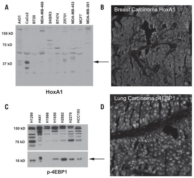
Not all antibodies that perform well in Western blot or ELISA are validated for immunohistochemistry.
What to check
- Has the antibody been validated for IHC?
- Is it compatible with formalin-fixed paraffin-embedded (FFPE) or frozen tissue?
- Was the recommended dilution used?
Solutions
- Use antibodies validated specifically for IHC applications.
- Optimize antibody concentration through serial dilution.
- Extend primary antibody incubation if necessary, such as overnight at 4°C.
- Include a known positive tissue as a control.
Possible Cause 3: Low or Absent Target Protein Expression
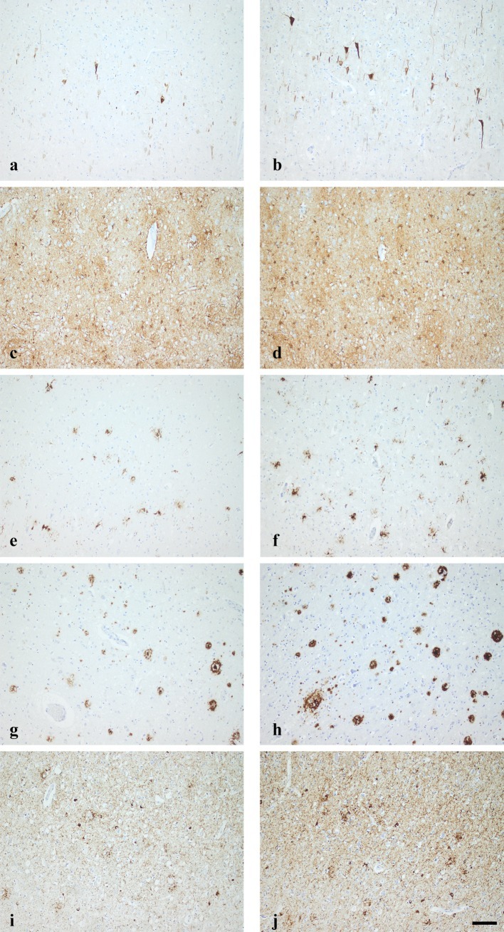
Sometimes the experiment is technically successful, but the selected tissue simply expresses little or no target protein.
What to check
- Is the target protein known to be expressed in the selected tissue?
- Is expression dependent on disease state, developmental stage, or treatment conditions?
Solutions
- Confirm expression using published literature or expression databases.
- Include a tissue with confirmed target expression as a positive control.
- Verify antibody performance using cultured cells or recombinant protein if available.
Possible Cause 4: Over-Fixation Reduced Antigen Accessibility
Excessive fixation can make antigen retrieval much more difficult.
What to check
- How long was the tissue fixed?
- Was fixation performed using neutral buffered formalin?
Solutions
- For most tissues, limit fixation to approximately 24 hours unless longer fixation is specifically required.
- Standardize fixation conditions across experiments.
- If over-fixation is unavoidable, optimize antigen retrieval conditions accordingly.
Possible Cause 5: Detection System Failure
If the secondary antibody or chromogenic detection reagents are not functioning properly, no signal will be produced even when antigen-antibody binding occurs.
What to check
- Is the secondary antibody compatible with the primary antibody species?
- Have the detection reagents expired?
- Was the DAB substrate freshly prepared?
Solutions
- Verify that the secondary antibody matches the host species of the primary antibody.
- Prepare fresh chromogenic substrate before use.
- Confirm the detection system using a positive control slide.
Possible Cause 6: Tissue Sections Lost Antigen Integrity
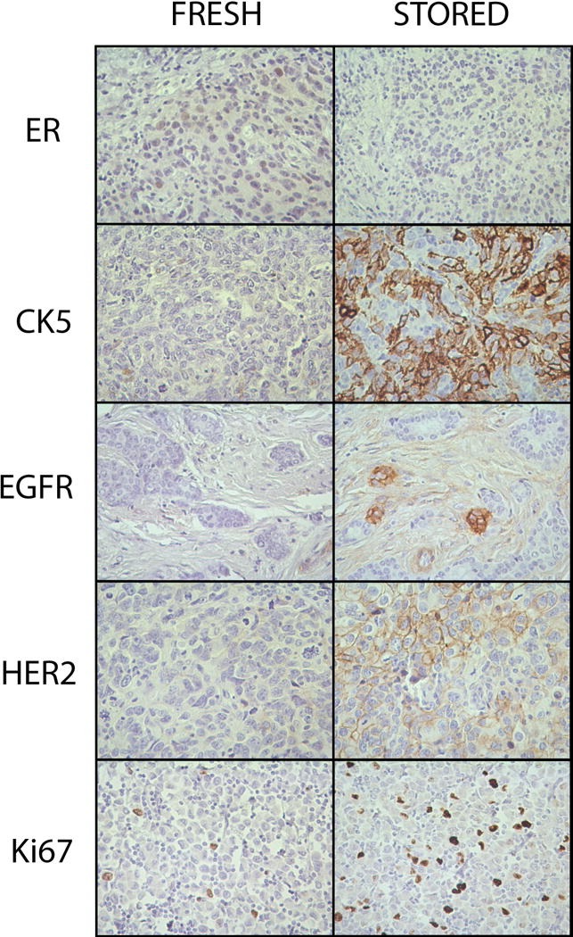
Improper tissue storage or repeated section handling can reduce antigen stability.
What to check
- How long have the slides been stored?
- Were tissue sections exposed to moisture or repeated temperature fluctuations?
Solutions
- Store unstained slides in a dry environment at appropriate temperature.
- Use freshly cut sections whenever possible.
- Avoid prolonged storage before staining.
Quick Troubleshooting Checklist
| Observation | Most Likely Cause | Recommended Action |
|---|---|---|
| No staining in all samples | Detection system failure | Check secondary antibody and chromogen |
| Positive control is also negative | Antigen retrieval or antibody issue | Optimize retrieval and verify antibody |
| Tissue morphology is good but no signal | Low antigen accessibility | Improve antigen retrieval conditions |
| Frozen tissue stains but FFPE does not | Epitope masking | Adjust retrieval buffer or method |
| Only weak staining observed | Low antibody sensitivity | Increase antibody concentration or incubation time |
Best Practices for Successful IHC
Reliable immunohistochemistry depends on consistent optimization throughout the workflow.
To improve staining success:
- Use tissues with confirmed target expression as positive controls.
- Standardize fixation and processing procedures.
- Optimize antigen retrieval for each target protein.
- Use antibodies validated for IHC.
- Include negative controls to evaluate non-specific staining.
- Prepare detection reagents fresh and verify their performance regularly.
Careful optimization at each step will significantly improve staining consistency and reproducibility.
Frequently Asked Question
Q: My Western blot shows a strong band, but my IHC staining is completely negative. Why?
Western blot detects denatured proteins after electrophoresis, whereas IHC detects proteins in their native tissue context. Formalin fixation can mask epitopes, making them inaccessible to certain antibodies. As a result, an antibody that performs well in Western blot may not recognize the same target in formalin-fixed tissue. Using an antibody validated for IHC and optimizing antigen retrieval are often necessary to obtain successful staining.


