
On May 18, the government of the Democratic Republic of the Congo (DRC) reported that the newly emerged Ebola outbreak in Ituri Province continues to spread. Authorities have recorded 116 suspected deaths, while another 435 suspected cases remain under observation and treatment.
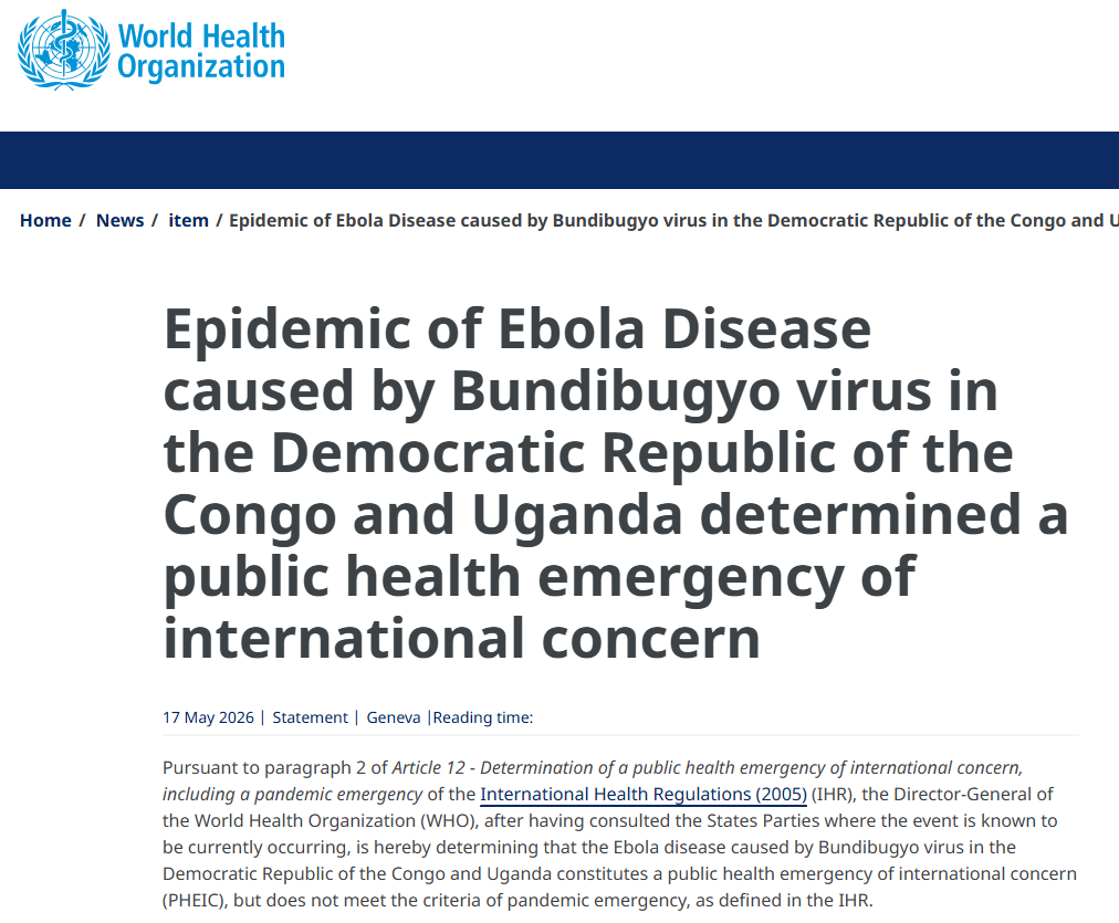
Meanwhile, neighboring Uganda has also reported a fatal imported case. The World Health Organization (WHO) has declared the outbreaks in the DRC and Uganda a "Public Health Emergency of International Concern (PHEIC)." Rwanda, Tanzania, Zambia, and other neighboring countries have subsequently strengthened border screening and public health surveillance measures.
At a time when the global public health system is still recovering from the aftermath of COVID-19, the resurgence of Ebola virus — one of the world's most lethal pathogens — has once again drawn international attention.
Ebola Virus: A Highly Lethal Filovirus
Ebola virus belongs to the genus Ebolavirus within the family Filoviridae. It is an enveloped, single-stranded negative-sense RNA virus. Viral particles typically appear as elongated filaments, although "U"-shaped, circular, and branched morphologies are also observed. Virions may extend several hundred to over one thousand nanometers in length.
The viral genome is approximately 19 kb long and encodes seven major structural proteins, including:
- NP (nucleoprotein), which encapsidates the viral RNA genome;
- GP (glycoprotein), the surface envelope protein responsible for host cell attachment and membrane fusion;
- VP24, VP35, and VP40, which participate in viral replication, assembly, and immune evasion;
- L protein, the RNA-dependent RNA polymerase required for viral transcription and genome replication.
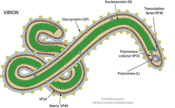
Schematic of the Ebolavirus structure
Among these proteins, GP glycoprotein is considered one of the key virulence determinants of Ebola virus and serves as the primary target for vaccine and neutralizing antibody development. Following receptor binding mediated by GP, the virus enters host cells and initiates intracellular replication and assembly.
Bundibugyo Ebolavirus: The Key Lineage Behind the Current Outbreak
According to publicly available data, the ongoing outbreak involves Bundibugyo ebolavirus, one of the six currently recognized Ebolavirus species. It is also one of the three Ebola virus species known to cause large-scale human outbreaks, alongside Zaire ebolavirus and Sudan ebolavirus.
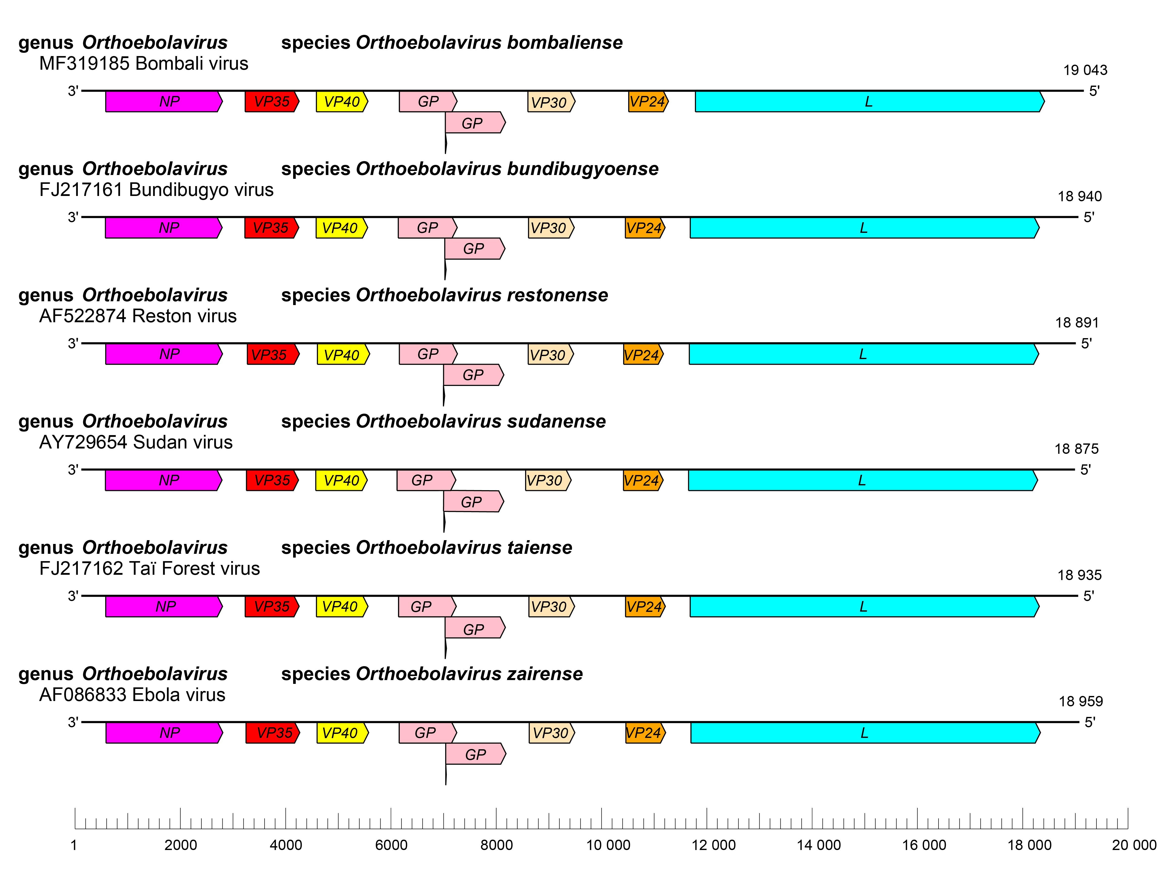
Schematic representation of orthoebolavirus genome organization. Genomes are drawn to scale (ICTV)
Compared with the Zaire species, Bundibugyo ebolavirus has caused fewer recorded outbreaks historically, but it remains highly pathogenic and transmissible. The virus was first identified in 2007 in the Bundibugyo district of Uganda during a major outbreak event.
Antigenic differences among Ebola virus species remain one of the major challenges in vaccine and therapeutic antibody development. Many currently approved vaccines were designed primarily against Zaire ebolavirus, and their protective efficacy against other species may be significantly reduced.
Transmission and Pathogenesis
Ebola virus is a classic zoonotic pathogen. Fruit bats are widely considered one of its primary natural reservoirs, and spillover transmission from wildlife to humans is believed to initiate outbreaks.
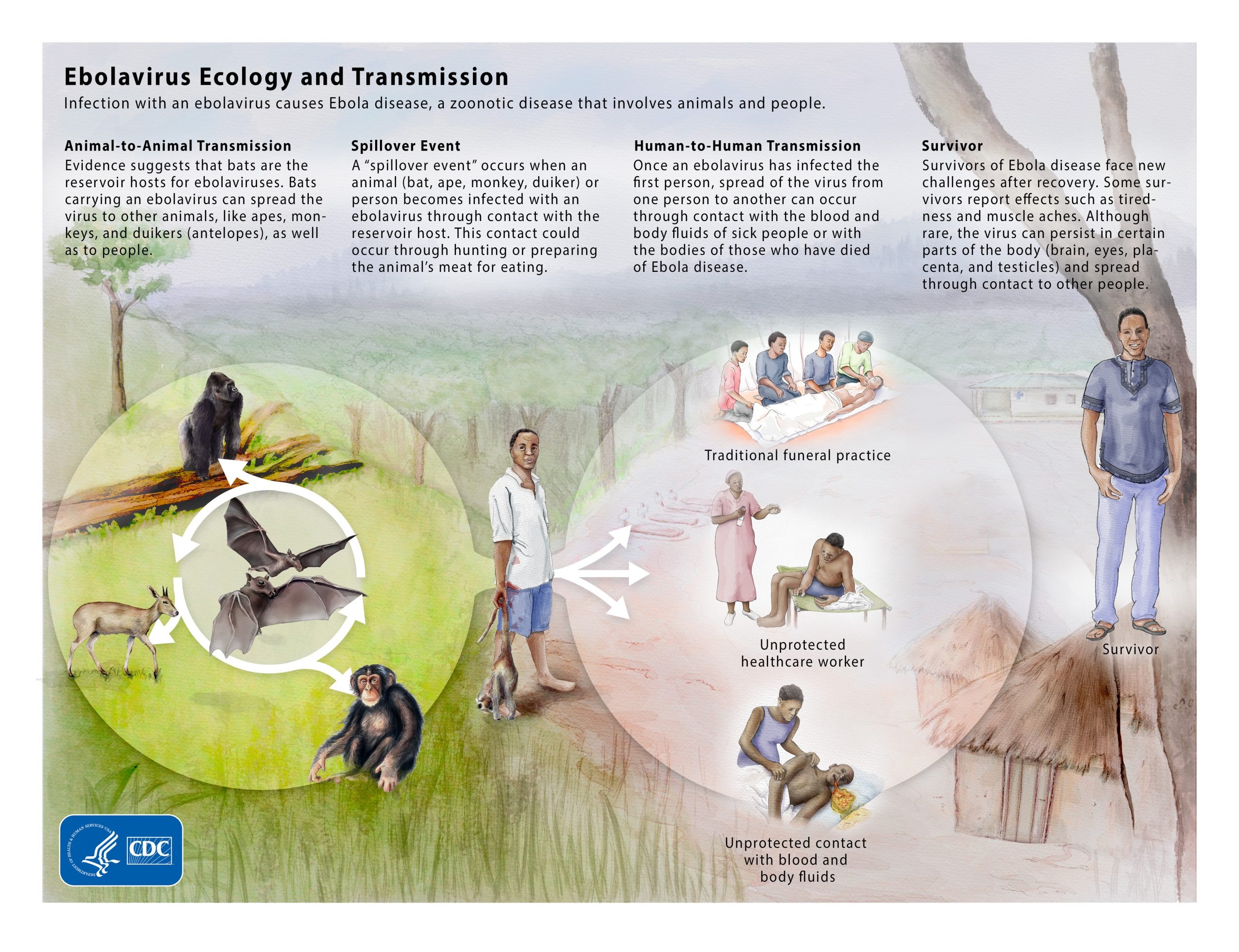
Ebolavirus Ecology and Transmission
Human infection typically occurs through direct contact with infected animals or exposure to the blood, body fluids, secretions, or tissues of infected individuals. Healthcare-associated transmission through contaminated medical equipment and close-contact exposure during funeral practices are also well-documented routes of spread.
Unlike influenza viruses or SARS-CoV-2, Ebola virus is not considered a typical airborne pathogen. Transmission largely depends on close physical contact. However, because viral loads in patient body fluids can become extremely high, outbreaks can spread rapidly in healthcare settings and household clusters, especially in regions with limited medical infrastructure.
Following infection, Ebola virus can extensively target monocytes, macrophages, dendritic cells, and vascular endothelial cells, triggering severe inflammatory responses, coagulation abnormalities, and vascular leakage. In severe cases, this may progress to multiorgan failure, shock, and hemorrhagic manifestations.
Viral Entry and Replication Mechanism
Ebola virus entry into host cells is highly dependent on its surface glycoprotein (GP). The virus initially attaches to the host cell membrane through GP and is subsequently internalized via macropinocytosis. Following uptake into endosomes, host cathepsins cleave GP, exposing critical binding regions that interact with the endosomal receptor Niemann–Pick C1 (NPC1). The interaction between GP and NPC1 triggers fusion between the viral envelope and endosomal membrane, releasing the viral nucleocapsid into the cytoplasm.
Once released, the negative-sense RNA genome undergoes primary transcription mediated by the Ebola virus polymerase complex, which consists of L, NP, VP35, and VP30. This process generates viral mRNAs that direct the synthesis of viral proteins. As viral proteins accumulate, NP and VP35 promote the formation of specialized replication compartments known as inclusion bodies (IBs), where viral genome replication predominantly occurs.
During replication, the negative-sense genomic RNA is first copied into a full-length positive-sense intermediate, which subsequently serves as the template for the synthesis of additional viral genomes. Newly assembled nucleocapsids then migrate to the periphery of inclusion bodies and are transported to the plasma membrane for viral budding. Meanwhile, GP is incorporated into the viral envelope, ultimately leading to the release of mature virions from the host cell surface.
In addition to its role in viral RNA synthesis, VP35 is also involved in nucleocapsid assembly, intracellular transport, and suppression of host innate immune responses, making it one of the central regulatory proteins throughout the Ebola virus life cycle.
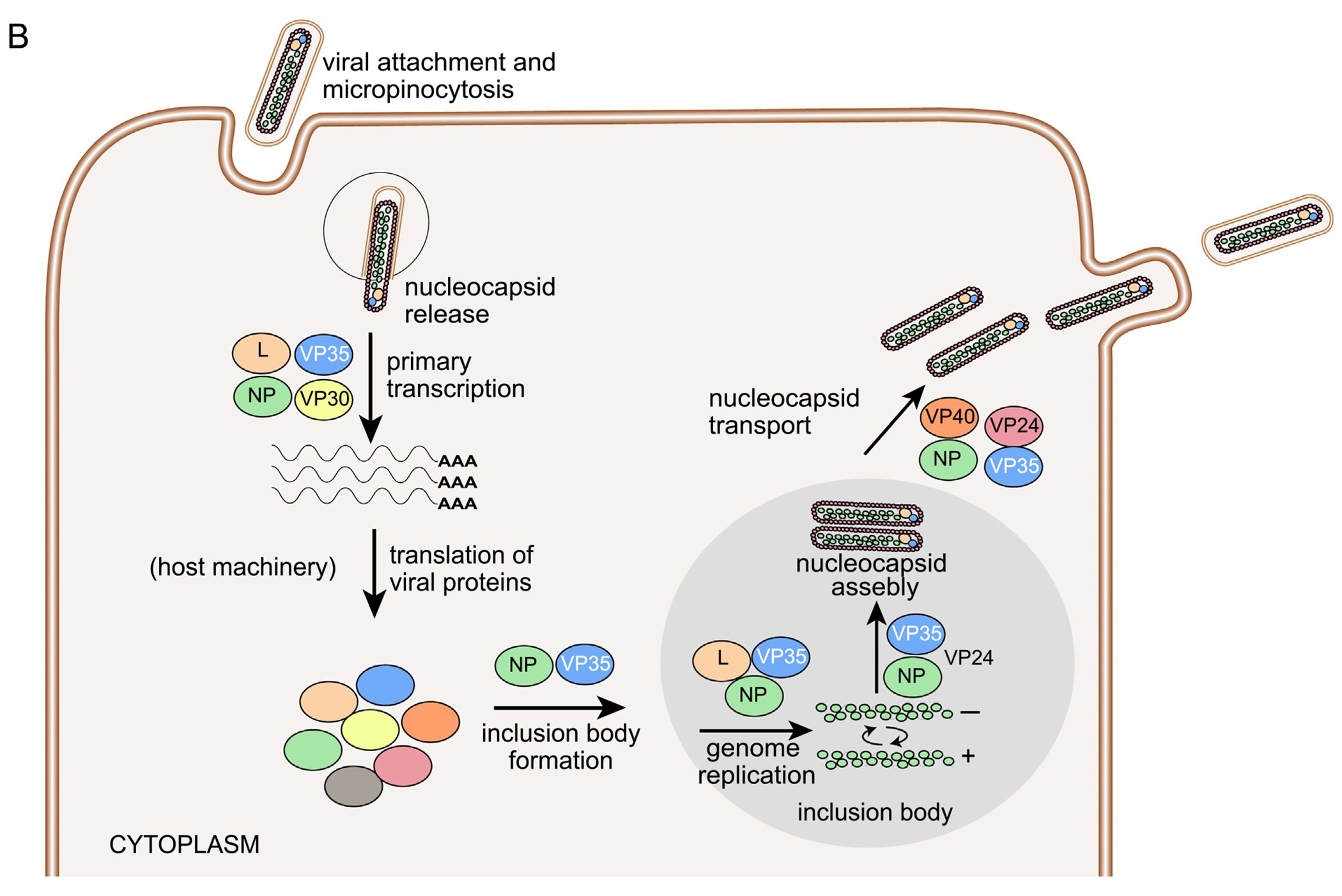
Ebola virus genome and infectious cycle (PMCID: PMC13140081)
Why Is Ebola So Deadly?
Ebola virus disease (EVD) typically has an incubation period ranging from 2 to 21 days.
Early symptoms are often nonspecific and resemble common viral infections, including:
- Fever
- Fatigue
- Muscle pain
- Headache
- Sore throat
As disease progression accelerates, patients may develop:
- Severe diarrhea and vomiting
- Liver and kidney dysfunction
- Coagulation abnormalities
- Internal and external bleeding
- Hypotensive shock
Depending on the viral species and outbreak conditions, case fatality rates may range from approximately 50% to as high as 90%.
Importantly, Ebola-associated mortality is not caused solely by hemorrhage itself. The virus simultaneously induces:
1. Severe inflammatory dysregulation;
2. Endothelial damage;
3. Coagulation dysfunction;
4. Multiorgan failure.
In regions lacking adequate intensive care capacity and supportive treatment, mortality risk increases substantially.
Why Do Outbreaks Persist Despite Approved Vaccines?
To date, three Ebola vaccines have been approved globally:
- Ervebo, developed by Merck & Co.;
- Zabdeno, developed by Johnson & Johnson;
- Ad5-EBOV, developed by CanSino Biologics.
However, vaccines have not eliminated Ebola outbreaks as effectively as many people expected, for several important reasons.
First, most currently available vaccines were designed primarily against Zaire ebolavirus. Antigenic differences between Ebola virus species mean that cross-protective immunity against Bundibugyo or Sudan species may be limited.
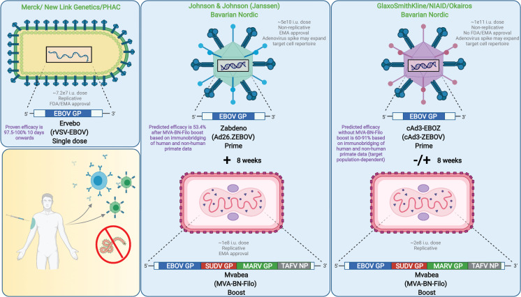
Leading EBOV vaccines (PMCID: PMC8659338)
Second, Ebola outbreaks frequently occur in regions with fragile healthcare systems and limited infrastructure. Vaccination campaigns often face substantial logistical and operational challenges, including cold-chain transportation constraints, insufficient healthcare resources, cross-border population movement, armed conflict, and vaccine hesitancy within local communities.
These factors can severely reduce vaccination efficiency and outbreak containment capacity.
In addition, Ebola outbreaks are often sudden, geographically localized, and difficult to predict. By the time vaccination programs are fully deployed, transmission chains may already be well established. Compared with seasonal vaccines for influenza or COVID-19, Ebola vaccines also face relatively limited long-term commercial demand, which affects manufacturing scale and global stockpile capacity.
Most importantly, there is still no broadly protective pan-Ebolavirus vaccine capable of covering multiple pathogenic Ebola species simultaneously. Developing broadly protective vaccines remains one of the central goals in current Ebola research.
Current Treatment Strategies
At present, supportive care remains the cornerstone of Ebola virus disease management, including:
- Fluid and electrolyte replacement;
- Respiratory support;
- Blood pressure stabilization;
- Renal support therapy;
- Coagulation monitoring and management.
In recent years, monoclonal antibody therapies targeting Zaire ebolavirus have shown encouraging clinical benefits. However, broadly effective therapeutics against multiple Ebola virus species remain limited.
As a result, continued research into viral biology, neutralizing antibodies, vaccine platforms, and rapid diagnostics remains critically important.
Conclusion
From the devastating West African epidemic to the newly expanding outbreaks in the DRC and Uganda, Ebola virus continues to represent a major global public health threat.
In an increasingly interconnected world, localized outbreaks can rapidly become international concerns. Strengthening viral surveillance, vaccine preparedness, cross-border coordination, and rapid diagnostic capacity will remain essential components of future global health security.
At the same time, the antigenic diversity among Ebola virus species continues to drive demand for broadly protective vaccines, neutralizing antibodies, and next-generation diagnostic technologies. Building a truly comprehensive defense system against highly lethal filoviruses remains one of the major scientific and public health challenges ahead.
Antigen
| Catalog | Product Name |
|---|---|
| EVV03605 | Recombinant REBOV GP1 Protein, C-Fc |
| EVV03604 | Recombinant REBOV GP1 Protein, C-His |
| YVV03602 | Recombinant REBOV GP1 Protein, N-His |
| EVV03607 | Recombinant SEBOV GP1 Protein, C-Fc |
| EVV03606 | Recombinant SEBOV GP1 Protein, C-His |
| YVV03603 | Recombinant SEBOV GP1 Protein, N-His |
| EVV03609 | Recombinant TAFV GP1 Protein, C-Fc |
| EVV03608 | Recombinant TAFV GP1 Protein, C-His |
| YVV03604 | Recombinant TAFV GP1 Protein, N-His |
| EVV03601 | Recombinant ZEBOV GP/GP1,2 Protein, C-His |
| EVV03603 | Recombinant ZEBOV GP1 Protein, C-Fc |
| EVV03602 | Recombinant ZEBOV GP1 Protein, C-His |
| YVV03601 | Recombinant ZEBOV GP1 Protein, N-His |
Antibody
| Catalog | Product Name |
|---|---|
| RVV24306 | Anti-pan-Filovirus GP/Envelope glycoprotein Antibody (m21D10) |
| PVV03602 | Anti-REBOV GP1 Polyclonal Antibody |
| RVV03603 | Anti-SEBOV GP/Envelope glycoprotein Antibody (16F6) |
| PVV03603 | Anti-SEBOV GP1 Polyclonal Antibody |
| RVV23301 | Anti-Sudan ebolavirus/SEBOV NP/Nucleoprotein Antibody (SAA1402) |
| PVV03604 | Anti-TAFV GP1 Polyclonal Antibody |
| RVV03601 | Anti-Zaire ebolavirus/ZEBOV GP/GP1,2 Nanobody (SAA1248) |
| RVV03604 | Anti-ZEBOV GP/Envelope glycoprotein Antibody (GPE118) |
| RVV03605 | Anti-ZEBOV GP/Envelope glycoprotein Antibody (KZ52) |
| RVV03602 | Anti-ZEBOV GP/Envelope glycoprotein Antibody (mAb100) |
| PVV03601 | Anti-ZEBOV GP1 Polyclonal Antibody |
| RVV23302 | Anti-ZEBOV NP/Nucleoprotein Antibody (KZ51) |
| RVV28501 | Anti-ZEBOV VP35/Polymerase cofactor VP35 Antibody (F9) |
| RVV28601 | Anti-ZEBOV VP40/Matrix protein VP40 Antibody (DSTL094) |
| VVV23301 | InVivoMAb Anti-REBOV/SEBOV/TAFV/ZEBOV NP/Nucleoprotein Antibody (MJ20) |
| VVV03605 | InVivoMAb Anti-SEBOV/ZEBOV GP/Envelope glycoprotein Antibody (Iv0198) |
| VVV03604 | InVivoMAb Anti-ZEBOV GP/Envelope glycoprotein Antibody (6D8) |
| DVV03610 | Research Grade Anti-REBOV/SEBOV/TAFV/ZEBOV GP/Envelope glycoprotein Antibody (ADI-15878) |
| DVV03608 | Research Grade Anti-ZEBOV GP/Glycoprotein (ANP-015) |
| DVV03609 | Research Grade Anti-ZEBOV GP/Glycoprotein (Zmapp) |


