
Please ensure Javascript is enabled for purposes of website accessibility
Home / Products / Super-Affinity Antibody

Anti-Mouse APOE Antibody (HJ6.3)

Catalog #:   RMB98601 Specific References (0) DATASHEET
Host species: Mouse
Isotype: IgG1, kappa
Applications: Blocking, IHC
Accession: P08226
Overview

Catalog No.

RMB98601

Species reactivity

Mouse

Host species

Mouse

Isotype

IgG1, kappa

Clonality

Monoclonal

Applications

Blocking, IHC

Target

Apo-E, Apolipoprotein E, APOE

Concentration

2.73 mg/ml

Endotoxin level

Please contact with the lab for this information.

Purity

>95% as determined by SDS-PAGE.

Purification

Protein A/G purified from cell culture supernatant.

Accession

P08226

Form

Liquid

Storage buffer

100 mM Pro-Ac, 20 mM Arg, pH 5.0

Stability and Storage

Use a manual defrost freezer and avoid repeated freeze-thaw cycles. Store at 4°C short term (1-2 weeks). Store at -20°C 12 months. Store at -80°C long term.

Clone ID

HJ6.3

Note

For research use only.

Data Image
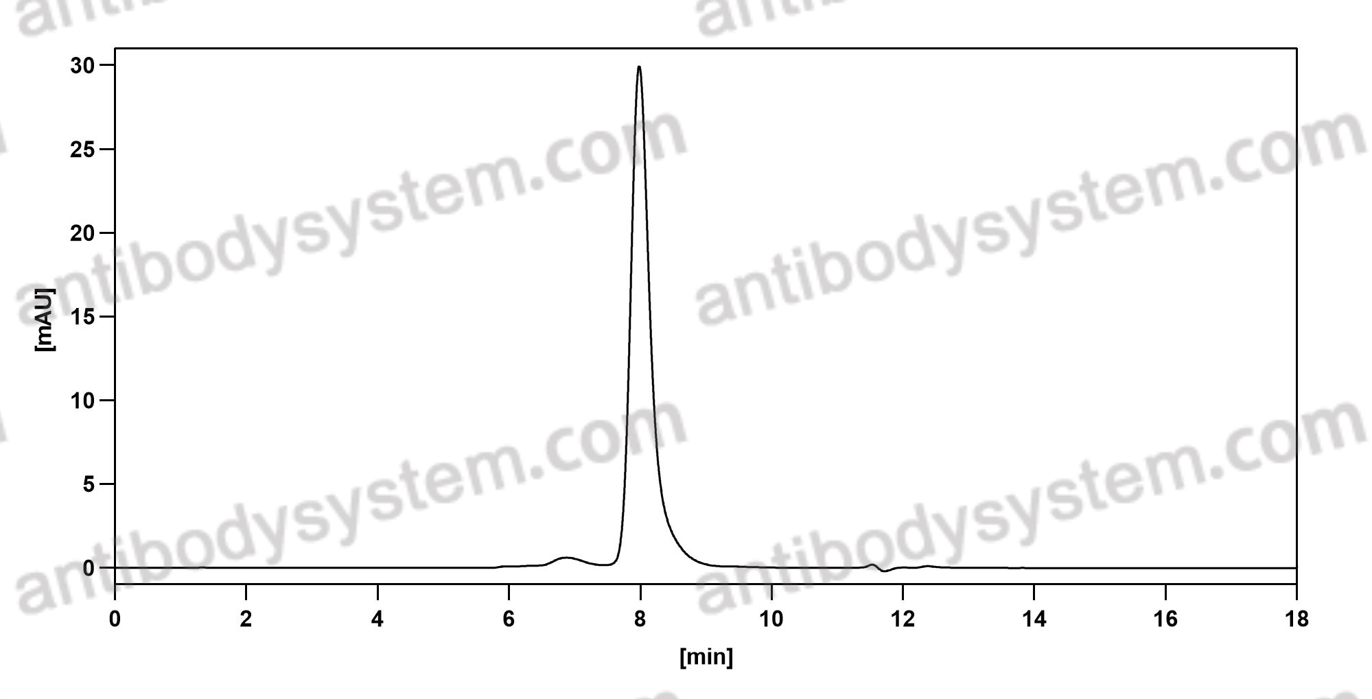
  • SEC-HPLC
    The purity of this product is >95% as determined by SEC-HPLC.
  • Bioactivity
    Fig. 7 APOE and PD-1 antibodies boost anti-tumor immunity in mouse GBM model. a Survival curves of C57B6/J mice bearing Trp53- mutated GL261 GBM treated with the indicated antibodies. The Mantel-Cox test is used for the significance calculation. c Representative IHC staining image showing APOE levels of CD8+ T cells in necrotic regions in GL261 GBM. d UMAP plot for the single-cell transcriptomic characterization of all cells in GBM from four treatment groups in a. e UMAP plot of CD8+ T cell subclusters and their proportions in the four groups of mouse GBM. f Transcriptomic comparisons of enriched signaling pathways and top feature genes expressed in CD8+ T cells from the groups of mouse GBM. g A working model to explain the effect of anti-APOE antibody in GBM treatment. Necrotic tumor cells release APOE lipid particles into the tumor microenvironment, which are taken up by infiltrating T cells. Lipid-laden T cells become dysfunctional and apoptotic. The APOE antibody reduces T cell apoptosis by neutralizing APOE lipid particles in the tumor microenvironment.
    Data sourced from citation (PMID: 40903538)
  • Western Blot
    Figure 5. APOE promotes inflammatory reactivity in microglia. C) Levels of the cytokines TNF𝛼, IL-1𝛽, and IL-6 in the conditioned medium. E) Western blot analysis displaying protein expression of TLR4, cGAS, phosphorylated-STING (p-STING), STING, and APOE in microglia isolated from PBS- or KA-treated groups, with 𝛽-actin as a loading control. F) Quantification of the relative expression of TLR4, cGAS, p-STING, STING, and APOE normalized to 𝛽-actin based on the Western blot data. G) A proposed model illustrating the potential mechanism by which APOE enhances inflammatory activation in microglia. Created by BioRender.com. All experiments were independently repeated three times.
    Data sourced from citation (PMID: 41085045)
References
Datasheet
$ 328
Product specifications
100 μg 328 1 mg 1640

Contact Information

Order: order@antibodysystem.com

Mail: support@antibodysystem.com

Distributor list

For research use only. Not for human or drug use.

Need help with your order?

Find out more about placing an order here

Anti-Mouse APOE Antibody (HJ6.3) [RMB98601]
Terms of sale Website terms of use Cookie policy Privacy
Copyright © 2025 AntibodySystem SAS. All Rights Reserved.            All Products are for Research Use Only