
Please ensure Javascript is enabled for purposes of website accessibility
Home / Products / Antibody / Recombinant Antibody

Anti-DNA-RNA Hybrid Antibody (S9.6)

Catalog #:   RGK60001 Specific References (0) DATASHEET
Host species: mouse
Isotype: IgG2a, kappa
Applications: CHIP, ChIP-seq, DB, EMSA, FISH, ICC, IF, IP, SPR
Expression system: Mammalian Cells
Overview

Catalog No.

RGK60001

Description

DNA-RNA hybrids are a natural occurrence within eukaryotic cells and their level are high at sites of high transcriptional activity. They are non-canonical nucleic acid structures with transcriptional regulatory functions. Their presence is reported to predispose a locus to chromosomal breakage. A locus forming an DNA:RNA creates a double-stranded A/B intermediate conformation, with a second target for single-stranded nucleic acid binding proteins on the complementary, displaced DNA strand. They are shown to be resistant to the activity of DNA methyltransferases. The formation of DNA:RNA hybrids has been associated with a number of neurological diseases. Mutations in the DNA:RNA helicase senataxin (SETX) are implicated in the dominant juvenile form of amyotrophic lateral sclerosis type 4 and a recessive form of ataxia oculomotor apraxia type 2.

Expression system

Mammalian Cells

Species reactivity

General

Host species

mouse

Isotype

IgG2a, kappa

Clonality

Monoclonal

Applications

CHIP, ChIP-seq, DB, EMSA, FISH, ICC, IF, IP, SPR

Target

DNA-RNA Hybrid

Concentration

3.82 mg/ml

Endotoxin level

Please contact with the lab for this information.

Purity

>95% as determined by SDS-PAGE.

Purification

Protein A/G purified from cell culture supernatant.

Form

Liquid

Storage buffer

100 mM Pro-Ac, 20 mM Arg, pH 5.0

Stability and Storage

Use a manual defrost freezer and avoid repeated freeze-thaw cycles. Store at 4°C short term (1-2 weeks). Store at -20°C 12 months. Store at -80°C long term.

Clone ID

S9.6

Note

For research use only.

Data Image
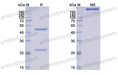
  • SDS-PAGE
    SDS PAGE for DNA-RNA Hybrid Antibody(S9.6)
  • Conjugation
    (a) SEM image and size distribution plot of WSNs. The scale bar is 200 nm. (b) TEM image of WSNs. The scale bar is 200nm. (c) ζ-Potential assay of surface modification of WSNs. (d) UV−vis absorption spectra of WSNs, Ab, and WSNs@Ab.
    Data sourced from citation (PMID: 38478499)
  • Immunofluorescence
    Figure 5. RNASEH1-R-loop signaling mediates PVC NPs-induced STING pathway activation in macrophages. A) Representative composite fluorescence images showing the levels of R-loops after treatment with different concentrations of PVC NPs. The R-loop and nuclei were labeled with an anti-DNA: RNA hybrid antibody (S9.6) (green) and DAPI (blue), respectively (scale bar = 5 μm). B) Quantitative analysis of the relative fluorescence intensity of R-loops in the nucleus of cells treated with different concentrations of PVC NPs (n > 50 cells). F) Representative R-loop PLA fluorescence images demonstrating the RNASEH1-R-loop interaction (scale bar = 5 μm). G) Quantitative analysis of PLA foci in the nuclear compartment (n > 50 cells). H) Representative composite fluorescence images showing the levels of R-loops after RNASEH1 overexpression following PVC NPs treatment (scale bar = 3 μm).
    Data sourced from citation (PMID: 40919700)
  • Western Blot
    Western blot analysis for RNA:DNA hybrid accumulation and DNA damage in 54 hpf embryos. A S9.6 antibody detected higher RNA:DNA hybrid levels in prpf4−/− embryos than in siblings.
    Data sourced from citation (PMID: 41360772)
  • Bioactivity
    (b) Fluorescence emission spectra of bQDs, bQDs-ssDNA1, and WSNs@Ab/bQDs-ssDNA1/miR-135b.
    Data sourced from citation (PMID: 38478499)
  • Bioactivity
    Optimization reaction conditions. (a)WSNs to S9.6 Ab mass ratio, (b) BSA concentration, and (c) QDs-ssDNA amount. As shown in Figure 4a, the fluorescence intensity increased with the ratios of WSNs to S9.6 Ab at 1:1,1:4, 1:7, and 1:10 and tended to be constant or even decreased along with the ratios more than 1:13 and 1:16. This indicated that Ab modified on the surface of WSNs reached saturation when the ratio was 1:10. The subsequent decrease may be due to the dense distribution of Ab causing steric hindrance to the capture of DNA/miRNA hybrids. To reduce the nonspecific adsorption of WSNs@Ab, we blocked the superficial active sites with a BSA-PBS solution. As shown in Figure 4b, the signal-to-background fluorescence ratio (F/F0, where F and F0 were the fluorescence intensities from the samples with and without miRNA, respectively) increased with BSA concentration and gained the highest signal at 0.25%. Insufficient concentration of BSA caused significant nonspecific adsorption of WSNs@Ab. Conversely, an excess concentration of BSA would adversely impact the Ab capture of hybrids. Moreover, the amount of QDs-ssDNA influenced the performance of miRNA detection. As shown in Figure 4c, the optimal amount was considered as 20 μL, as the fluorescence intensity reached a plateau at this volume. Thus, the mass ratio of WSNs to S9.6 Ab was set at 1:10, and 0.25% BSA and 20 μL of QDs-ssDNA were chosen for subsequent experiments.
    Data sourced from citation (PMID: 38478499)
References
Datasheet
$ 372
Product specifications
100 μg 372 1 mg 1860

Contact Information

Order: order@antibodysystem.com

Mail: support@antibodysystem.com

Distributor list

For research use only. Not for human or drug use.

Need help with your order?

Find out more about placing an order here

Anti-DNA-RNA Hybrid Antibody (S9.6) [RGK60001]
Terms of sale Website terms of use Cookie policy Privacy
Copyright © 2025 AntibodySystem SAS. All Rights Reserved.            All Products are for Research Use Only