The Exosomes were first found in sheep reticulocytes in 1983. Johnstone et al tracked transferrin receptors during the maturation of reticulocytes and found that the formation of exosomes is the mechanism for the loss of transferrin receptors in mature red blood cells. To distinguish them from other types of extracellular vesicles (EVs), they were named exosomes.
Exosomes, with a diameter of about 40–100nm, are biological nanoscale spherical lipid bilayer vesicles secreted by cells, and exosomes contain nucleic acids, proteins, lipids, cytokines, transcription factor receptors and other bioactive substances. With the in-depth study of exosomes, its applications are becoming more and more widespread.
Exosomes can play a role in physiological and pathological processes, that plays important roles in many aspects of human health and disease, including development, immunity, tissue homeostasis, cancer, and neurodegenerative diseases. In addition, viruses co-opt exosome biogenesis pathways both for assembling infectious particles and for establishing host permissiveness. On the basis of these and other properties,exosomes are being developed as therapeutic agents in multiple disease models.
Almost all types of normal cells can produce exosomes, like human umbilical vein endothelial cells, mesenchymal stem cells (MSC), T cells, B cells, macrophages, dendritic cells (DC), natural killer (NK) cells. Also, exosomes mentioned above can be widespread in biofluids, such as saliva, plasma, urine, ascites, milk and bile. Tumor cells can secrete a large number of exosomes, and the specific antigens on their surface can reflect the nature of donor cells.

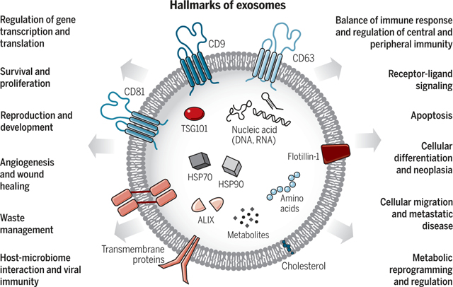
The structure of exosomes and their associated biological functions(Science. 2020 Feb 7;367(6478):eaau6977)
Exosome Characterization Methods
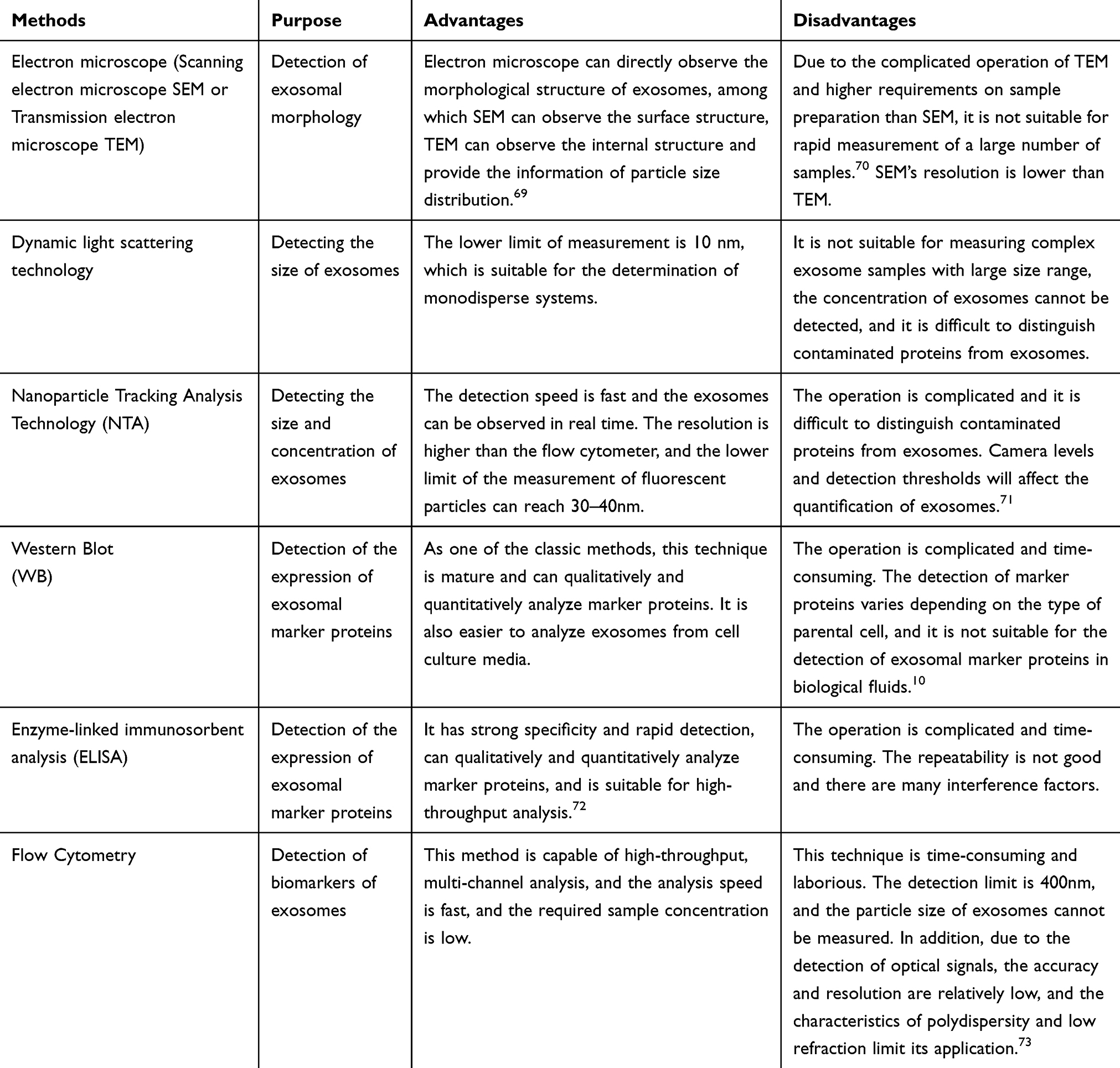
According to the International Society for Extracellular Vesicles (ISEV), three key criteria were established in 2014 for the identification of exosomes: transmission electron microscopy (TEM), nanoparticle tracking analysis (NTA), and detection of protein markers by Western blot (WB). Figure 3 illustrates the commonly used characterization techniques for exosomes.

Purpose, Advantages and Disadvantages of Common Exosome Characterization Methods(Int J Nanomedicine. 2020 Sep 22:15:6917-6934.)
Exosome biomarkers
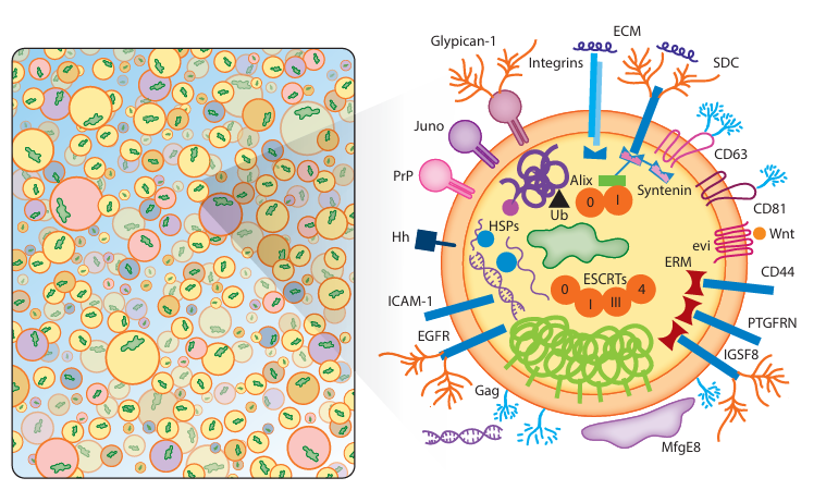
The protein composition of extracellular vesicles is a good indicator of the subtype of EV, the mode of biogenesis and release, and the original cell type. However, regardless of these factors, all exosomes contain some common proteins, for instance, heat shock protein 84 (Hsp84), tumor susceptibility gene 101 (TSG101) and Alix, for transport mechanism. The endosomal ESCRT is a collection of proteins required for the membrane infoldings to form MVBs. Five protein complexes comprise the ESCRT machinery, ESCRT-0, ESCRT-I, ESCRT-II, ESCRT-III and Vps4-Vta1, and a few ESCRT-associated proteins. Among them, the soluble complexes are ESCRT-0, ESCRT-I, ESCRT-II and Vps4-Vta1. ESCRT complex components, GTPases, Rab, tetraspanin, and other proteins play a role in the recognition, uptake, release, and exosome internalization. Alix, TSG101, Hsp70, and CD9 were the exosome markers used in the Western blotting and electron microscopy techniques. Other commonly found exosomal proteins include:
·Tetraspanins: CD9, CD81, CD82, Tetraspanin-8
·Heat shock proteins: Hsp84, HSP70, HSP90
·Receptors: CD46, CD55, NOTCH1
·Membrane adhesion proteins: Integrins
·Membrane transport/trafficking-related proteins: Annexins, Rab protein family
·Cytoskeletal components: Ezrin, Actin, Tubulin, Cytokeratins, Myosin
·Lysosomal marker proteins: Lysosome-associated membrane protein 1/2 (LAMP-1/2), Cathepsin D, CD63
·Antigen-presenting proteins: HLA class I and II/peptide complexes
·Metabolic enzymes: GAPDH, Pyruvate kinase
·Proteases: ADAM10, DPEP1, ST14
·Transporters: ATP7A, ATP7B, MRP2, SLC1A4, SLC16A1, CLIC1

Exosome biomarkers(Annu Rev Biochem. 2019 Jun 20:88:487-514)
Reference
1. Rezaie J, Feghhi M, Etemadi T. A review on exosomes application in clinical trials: perspective, questions, and challenges. Cell Communication and Signaling. 2022;20(1).
2. Xu G, Jin J, Fu Z, et al. Extracellular vesicle-based drug overview: research landscape, quality control and nonclinical evaluation strategies. Signal Transduction and Targeted Therapy. 2025;10(1).
3. Kurian TK, Banik S, Gopal D, Chakrabarti S, Mazumder N. Elucidating Methods for Isolation and Quantification of Exosomes: A Review. Molecular Biotechnology. 2021;63(4):249-266.
4. Pegtel DM, Gould SJ. Exosomes. Annual Review of Biochemistry. 2019;88(1):487-514.
5. Zhang Y, Bi J, Huang J, Tang Y, Du S, Li P. Exosome: A Review of Its Classification, Isolation Techniques, Storage, Diagnostic and Targeted Therapy Applications. International Journal of Nanomedicine. 2020;15:6917-6934.
AntibodySystem provides Exosomes-related proteins and antibodies, delivering more tools and solutions for research.
|
Catalog |
Product Name |
|
RHC69302 |
Anti-HSP70 Antibody (R1M49) |
|
RHE05802 |
Anti-HSPA4/HSP70 Antibody (R3J59) |
|
FHD48812 |
Anti-Human CD9 Antibody (SAA0003), PE |
|
RHD48802 |
Anti-Human CD9 Nanobody (SAA0905) |
|
DHD48801 |
Research Grade Anti-Human CD9 (AT14-012) |
|
VHD48801 |
InVivoMAb Anti-Human CD9 Antibody (Iv0274) |
|
RHC37602 |
Anti-CD63 Antibody (R3B43) |
|
RMC37601 |
Anti-CD63 Antibody (R1L87) |
|
FHF34010 |
Anti-Human CD81 Antibody (5A6) |
|
RHF34002 |
Anti-CD81 Antibody (R1Z70) |
|
RHJ47103 |
Anti-TSG101 Antibody (R3W11) |
|
VHJ47102 |
InVivoMAb Anti-Human TSG101 Antibody (Iv0254) |
|
RHJ47102 |
Anti-TSG101 Antibody (R2J91) |
|
PHJ29601 |
Anti-PDCD6IP Polyclonal Antibody |
|
RHJ29604 |
Anti-PDCD6IP/ALIX Antibody (R2J32) |
|
RHJ29603 |
Anti-PDCD6IP Antibody (R2J31) |
|
PHG16101 |
Anti-GM130/GOLGA2 Polyclonal Antibody |
|
RHG16103 |
Anti-GM130/GOLGA2 Antibody (R3R68) |
|
RHB37201 |
Anti-FLOT1 Antibody (R1B06) |
|
RMB37201 |
Anti-FLOT1 Antibody (R2V99) |
|
RHE80601 |
Anti-RAB27A Antibody (R3M45) |
|
RHF37503 |
Anti-ACTB/β-actin/Beta Actin Antibody (R3N80) |
|
RHC09001 |
Anti-GAPDH Antibody (R2Y76) |
